. 2020 Sep 1;60(3):581-593.
doi: 10.1093/icb/icaa099.

The Effects of Premature Tooth Extraction and Damage on Replacement Timing in the Green Iguana

Affiliations

The Effects of Premature Tooth Extraction and Damage on Replacement Timing in the Green Iguana

Kirstin S Brink et al. Integr Comp Biol. .

Abstract

Reptiles with continuous tooth replacement, or polyphyodonty, replace their teeth in predictable, well-timed waves in alternating tooth positions around the mouth. This process is thought to occur irrespective of tooth wear or breakage. In this study, we aimed to determine if damage to teeth and premature tooth extraction affects tooth replacement timing long-term in juvenile green iguanas (Iguana iguana). First, we examined normal tooth development histologically using a BrdU pulse-chase analysis to detect label-retaining cells in replacement teeth and dental tissues. Next, we performed tooth extraction experiments for characterization of dental tissues after functional tooth (FT) extraction, including proliferation and β-Catenin expression, for up to 12 weeks. We then compared these results to a newly analyzed historical dataset of X-rays collected up to 7 months after FT damage and extraction in the green iguana. Results show that proliferation in the dental and successional lamina (SL) does not change after extraction of the FT, and proliferation occurs in the SL only when a tooth differentiates. Damage to an FT crown does not affect the timing of the tooth replacement cycle, however, complete extraction shifts the replacement cycle ahead by 4 weeks by removing the need for resorption of the FT. These results suggest that traumatic FT loss affects the timing of the replacement cycle at that one position, which may have implications for tooth replacement patterning around the entire mouth.


Figures

Fig. 1
Fig. 1
Tooth anatomy and histology of I. iguana (color in supplementary files, B&W in print). (A) I. iguana skull in right lateral view, from Edmund’s original experiments, ROM R337. (B) Teeth of I. iguana, with resorption pits in functional teeth for replacement teeth (not preserved when soft tissue removed from skull, arrows). (C–C′) Histology of a tooth family with RT in late cap stage, resorbing FT. (D–D′) Histology of an FT with DL and RT with undifferentiated SL. (E–E′) β-Catenin staining. (F–F′) Tn-C staining. (G–G′) Staining of a 3-h BrdU pulse. (H–H′) Staining of a 1-week BrdU pulse labeling (Time 0, no chase) with positive cells (arrows) in the dl (dotted outline). (I–I′) Staining for BrdU after a 2-week chase. (J–J′) Staining for BrdU after an 8-week chase. (K) Diagram illustrates the distribution of TA cells (black dots) and LRCs (white dots) in the iguana DL. (L) Counts of BrdU-positive cells in the DL and SL at 1 week (no chase), 2 weeks chase, and 8 weeks chase. N = 3 teeth/timepoint. Scale bar: (A) 1 cm, (B) 1 mm, (C–J) 100 µM.
Fig. 2
Fig. 2
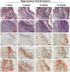
Differentiation of DL after FT extraction in I. iguana (Color in supplementary files, B&W in print). (A–L) Tooth regeneration 1 (A–C′), 3 (D–F′), 6 (G–I′), and 12 weeks (J–L) after FT extraction. (A–A′, D–D′, G–G′, and J–J′) H&E staining. There is no change in morphology in the SL until 12 weeks when it differentiates to a tooth bud. (B, E, H, and K) Staining of a 3-h BrdU pulse labeling. The SL does not have BrdU-positive cells after regenerating for 1(B), 3 (E), and 6 weeks, (H) however, cell proliferation is detected in the cervical loops of the developing RT. Cell proliferation is detected in the SL after 12 weeks of regeneration after differentiation of a new tooth (K, arrows). (C–C′ F–F′, I–I′ and L–L′) β-catenin staining. Nuclear β-catenin staining can be detected in the SL 1, 3, and 6 weeks after FT (arrows). β-catenin is diffusely present at 6 weeks. Nuclear β-catenin staining can be detected in the developing tooth bud at 12 weeks (arrows). Scale bar: 100 µM.
Fig. 3
Fig. 3
X-rays of tooth extraction and recovery in medial view in I. iguana. (A–A′′′) ROM R349, right maxilla. (A) before tooth extraction. Teeth 12–17 were removed during surgery (arrows). (A′) immediately after extraction. Teeth 12, 14, 15, and 17 were incompletely removed (asterisks). (A′′) 1-month post-extraction. Tooth 13 and tooth 16 erupted (arrows). (A′′′) 6 months post-extraction. Tooth position 15 suffered damage and did not return by the end of the experiment (arrow). (B–B′′′) ROM R309, left maxilla. (B) before tooth extraction. Teeth 12–15 were removed during surgery (arrows). (B′) 1-week post-extraction. Tooth 12 was incompletely removed (asterisk). (B′′) 2 months post-extraction. Tooth 13 erupted shorter than teeth in the rest of the toothrow (arrow). (B′′′) 7 months post-extraction. Tooth 13 has been shed and now has a normal phenotype. (C–C′′′) ROM R305, left mandible. (C) before tooth extraction. Teeth 10–13 were removed during surgery (arrows). (C′) 1-week post-extraction. Tooth 10 and 12 were incompletely removed (asterisks). (C′′) 3 months post-extraction. Tooth 11 erupted and ankylosed to the jaw at an angle (arrow). (C′′′) 6 months post-extraction. Teeth have normal phenotypes. Black dots are marks made by A.G. Edmund during preliminary analyses in the 1960s.
Fig. 4
Fig. 4
Timeline of tooth development in I. iguana. The time from shed to shed is ∼20 weeks in the animals examined in this study. (A) Normal development and development with damage to the crown. After an FT is shed, the RT erupts within a few weeks. The RT continues to develop and attach to the bone for another ∼8 weeks. Once it is fully attached, a new tooth is initiated in the SL at approximately 10–12 weeks. This new tooth then grows and resorbs the FT until it is shed. If the crown is removed, the base of the tooth still needs to be resorbed before the RT can erupt. (B) Shuffled timeline in the newly treated animals, where teeth were extracted when the SL had differentiated into a cap-stage tooth, ∼6 weeks before the FT would normally shed (Week 14 versus Week 20). Initiation of the next tooth begins 10–12 weeks after extraction, 4–5 weeks earlier than would be expected with normal shedding (Weeks 10–11, asterisk). The total replacement cycle is still 20 weeks, however, has been shifted ahead 4–5 weeks.

References

    1. An Z, Sabalic M, Bloomquist RF, Fowler TE, Streelman T, Sharpe PT.. 2018. A quiescent cell population replenishes mesenchymal stem cells to drive accelerated growth in mouse incisors. Nat Commun 9:378. - PMC - PubMed
    1. Berkovitz B, Moore M.. 1974. A longitudinal study of replacement patterns of teeth on the lower jaw and tongue in the rainbow trout Salmo gairdneri. Arch Oral Biol 19:1111–9. - PubMed
    1. Berkovitz B, Moore M.. 1975. Tooth replacement in the upper jaw of the rainbow trout (Salmo gairdneri). J Exp Zool 193:221–34. - PubMed
    1. Bertin TJC, Thivichon-Prince B, LeBlanc ARH, Caldwell MW, Viriot L.. 2018. Current perspectives on tooth implantation, attachment, and replacement in Amniota. Front Physiol 9:1630. - PMC - PubMed
    1. Brink K. 2020. Data accompanying: The effects of premature tooth extraction and damage on replacement timing in the green iguana.V1 ed. University of Manitoba 10.34990/FK2/SDWWBV). - DOI - PMC - PubMed

Publication types