. 2020 Dec;28(2):647-659.
doi: 10.1007/s40199-020-00372-7. Epub 2020 Sep 24.

Long-chain polyunsaturated omega-3 fatty acids reduce multiple myeloma exosome-mediated suppression of NK cell cytotoxicity

Affiliations

Long-chain polyunsaturated omega-3 fatty acids reduce multiple myeloma exosome-mediated suppression of NK cell cytotoxicity

Milad Moloudizargari et al. Daru. 2020 Dec.

Abstract

Background: Despite the advances in the treatment of multiple myeloma (MM), complete remission is usually challenging. The interactions between tumor and host cells, in which exosomes (EXs) play critical roles, have been shown to be among the major deteriorative tumor-promoting factors herein. Therefore, any endeavor to beneficially target these EX-mediated interactions could be of high importance.

Objectives: a) To investigate the effects of myeloma EXs on natural killer (NK) cell functions. b) To check whether treatment of myeloma cells with eicosapentaenoic acid (EPA) or docosahexaenoic acid (DHA), two polyunsaturated omega-3 fatty acids with known anti-cancer effects, can modify myeloma EXs in terms of their effects on natural killer functions.

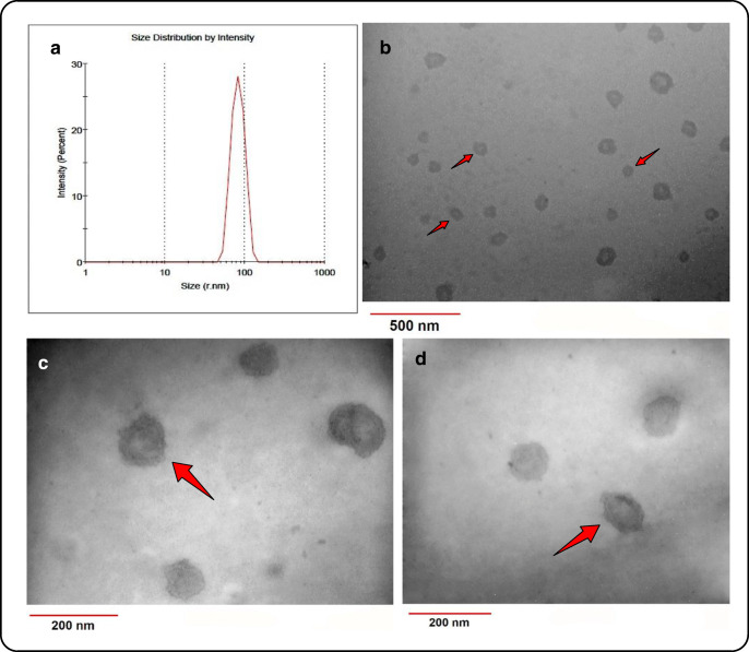
Methods: L363 cells were treated with either EPA or DHA or left untreated and the released EXs (designated as E-EX, D-EX and C-EX, respectively) were used to treat NK cells for functional studies.

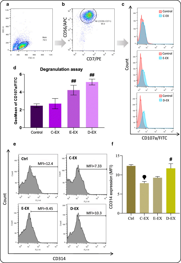
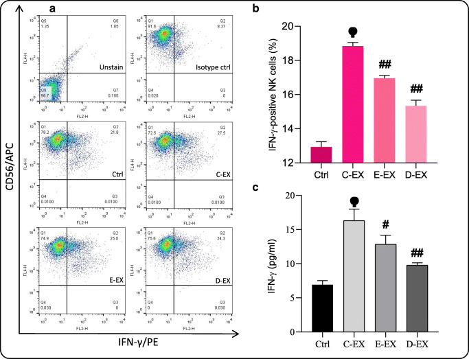
Results: Myeloma EXs (C-EXs) significantly reduced NK cytotoxicity against K562 cells (P ≤ 0.05), while the cytotoxicity suppression was significantly lower (P ≤ 0.05) in the (E-EX)- and (D-EX)-treated NK cells compared to the (C-EX)-treated cells. The expression of the activating NK receptor NKG2D and NK degranulation, after treatment with the EXs, were both altered following the same pattern. However, C-EXs could increase IFN-γ production in NK cells (P < 0.01), which was not significantly affected by EPA/DHA treatment. This indicates a dual effect of myeloma EXs on NK cells functions.

Conclusion: Our observations showed that myeloma EXs have both suppressive and stimulatory effects on different NK functions. Treatment of myeloma cells with EPA/DHA can reduce the suppressive effects of myeloma EXs while maintaining their stimulatory effects. These findings, together with the previous findings on the anti-cancer effects of EPA/DHA, provide stronger evidence for the repositioning of the currently existing EPA/DHA supplements to be used in the treatment of MM as an adjuvant treatment. EXs released from L363 (myeloma) cells in their steady state increase IFN-γ production of NK cells, while reduce their cytotoxicity against the K562 cell line (right blue trace). EXs from L363 cells pre-treated with either EPA or DHA are weaker stimulators of IFN-γ production. These EXs also increase NK cytotoxicity and NKG2D expression (left brown trace) compared to the EXs obtained from untreated L363 cells. Based on these findings, myeloma EXs have both suppressive and stimulatory effects on different NK functions depending on the properties of their cells of origin, which can be exploited in the treatment of myeloma.

Keywords: Cancer; Exosome; Extracellular vesicle; Natural killer cell; Omega-3; Tumor.


Figures

None
EXs released from L363 (myeloma) cells in their steady state increase IFN-γ production of NK cells, while reduce their cytotoxicity against the K562 cell line (right blue trace). EXs from L363 cells pre-treated with either EPA or DHA are weaker stimulators of IFN-γ production. These EXs also increase NK cytotoxicity and NKG2D expression (left brown trace) compared to the EXs obtained from untreated L363 cells. Based on these findings, myeloma EXs have both suppressive and stimulatory effects on different NK functions depending on the properties of their cells of origin, which can be exploited in the treatment of myeloma.
Fig. 1
Fig. 1
Illustrated flowchart of the methodology used in the study
Fig. 2
Fig. 2
Structural and size distribution analysis of the isolated MM-EXs. (A) Size distribution by intensity reveals a diameter range of 30 to 110 nm for the isolated EXs. (B, C and D) TEM images show membranous cup-shaped structures, the majority of which possess diameters of less than 100 nm
Fig. 3
Fig. 3
Flow cytometric characterization of EXs and protein content quantification. (A) reveals that the isolated EXs are positive for both CD81 and CD63. EPA and DHA treatment of the MM cells increased the percentage of particles with positive surface markers, which may be due to increased EX release from these cells. (B) BCA protein assay also shows increased protein content of the EXs after treatment with either EPA or DHA. ## Significantly different compared to the C-EX group (P < 0.01)
Fig. 4
Fig. 4
Cytotoxicity studies of NK cells. K562 cells were labeled with CFSE (5 μM) prior to their co-culture with NK-92 cells in each treatment group. (A and B) The CFSE+ population (target K562 cells) were initially gated. (C) After 4 h of co-culture, the cells were stained with PI and then subjected to flow cytometry analysis. In order to calculate the percentage of cytotoxicity, the percentage of PI+ cells were determined within the CFSE+ population, which were deemed as killed targets (%). (D) Shows the percentage of killed target cells as determined by flow cytometry. Each bar represents the Mean ± SEM of three different repeats. (E) Specific cytotoxicity of NK cells in different groups calculated based on the extent of LDH release. Each bar represents the Mean ± SEM of three different experiments. # significantly different compared to the C-EX group (P < 0.05) ## (P < 0.01) ϕ Significantly different compared to the control group (P < 0.01)
Fig. 5
Fig. 5
Flow cytometry studies of degranulation and CD314 expression. (A and B) NK-92 cells were identified using the markers CD56 and CD7. (C) Surface expression of CD107a as a degranulation marker was then detected using an anti-CD107a antibody. (D) The mean fluorescence intensity (MFI) of three independent experiments were then presented as bars. (E) Flow cytometry histograms of NKG2D expression in each treatment group. (F) C-EXs decreased NKG2D expression of NK-92 cells, while this effect was much weaker in the (E-EX)- and (D-EX)-treated groups. # Significantly different compared to the C-EX group (P < 0.05) ## (P < 0.01) ϕ Significantly different compared to the control group (P < 0.01)
Fig. 6
Fig. 6
IFN-γ production and release by NK-92 cells. (A and B) The percentage of IFN-γ positive NK cells was determined in each group following the intra-cellular staining. (C) The cytokine release of NK cells was then measured using ELISA. Based on the results, C-EX strongly stimulated IFN-γ production of NK cells. E-EX and D-EX also stimulated IFN-γ production, however the effect was not as pronounced as that of C-EX. # Significantly different compared to the C-EX group (P < 0.05) ## (P < 0.01) ϕ Significantly different compared to the control group (P < 0.01)

References

    1. Canella A, et al. The potential diagnostic power of extracellular vesicle analysis for multiple myeloma. Expert Rev Mol Diagn. 2016;16(3):277–284. - PubMed
    1. Iaccino E, et al. Monitoring multiple myeloma by idiotype-specific peptide binders of tumor-derived exosomes. Mol Cancer. 2017;16. - PMC - PubMed
    1. Di Marzo L, et al. Microenvironment drug resistance in multiple myeloma: emerging new players. Oncotarget. 2016;7(37):60698–60711. - PMC - PubMed
    1. Zhang L, Pan L, Xiang B, Zhu H, Wu Y, Chen M, Guan P, Zou X, Valencia CA, Dong B, Li J, Xie L, Ma H, Wang F, Dong T, Shuai X, Niu T, Liu T. Potential role of exosome-associated microRNA panels and in vivo environment to predict drug resistance for patients with multiple myeloma. Oncotarget. 2016;7(21):30876–30891. - PMC - PubMed
    1. Soley L, Falank C, Reagan MR. MicroRNA transfer between bone marrow adipose and multiple myeloma cells. Current Osteoporosis Reports. 2017;15(3):162–170. - PMC - PubMed

MeSH terms