C9orf72-Associated Arginine-Rich Dipeptide Repeat Proteins Reduce the Number of Golgi Outposts and Dendritic Branches in Drosophila Neurons
- PMID: 32975212
- PMCID: PMC7528685
- DOI: 10.14348/molcells.2020.0130
C9orf72-Associated Arginine-Rich Dipeptide Repeat Proteins Reduce the Number of Golgi Outposts and Dendritic Branches in Drosophila Neurons
Abstract
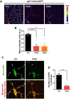
Altered dendritic morphology is frequently observed in various neurological disorders including amyotrophic lateral sclerosis (ALS) and frontotemporal dementia (FTD), but the cellular and molecular basis underlying these pathogenic dendritic abnormalities remains largely unclear. In this study, we investigated dendritic morphological defects caused by dipeptide repeat protein (DPR) toxicity associated with G4C2 expansion mutation of C9orf72 (the leading genetic cause of ALS and FTD) in Drosophila neurons and characterized the underlying pathogenic mechanisms. Among the five DPRs produced by repeat-associated non-ATG translation of G4C2 repeats, we found that arginine-rich DPRs (PR and GR) led to the most significant reduction in dendritic branches and plasma membrane (PM) supply in Class IV dendritic arborization (C4 da) neurons. Furthermore, expression of PR and GR reduced the number of Golgi outposts (GOPs) in dendrites. In Drosophila brains, expression of PR, but not GR, led to a significant reduction in the mRNA level of CrebA, a transcription factor regulating the formation of GOPs. Overexpressing CrebA in PR-expressing C4 da neurons mitigated PM supply defects and restored the number of GOPs, but the number of dendritic branches remained unchanged, suggesting that other molecules besides CrebA may be involved in dendritic branching. Taken together, our results provide valuable insight into the understanding of dendritic pathology associated with C9-ALS/FTD.
Keywords: C9orf72; CrebA; Golgi outposts; amyotrophic lateral sclerosis; dendrites.
Conflict of interest statement
The authors have no potential conflicts of interest to disclose.
Figures


Similar articles
-
C9orf72 Dipeptide Repeats Cause Selective Neurodegeneration and Cell-Autonomous Excitotoxicity in Drosophila Glutamatergic Neurons.J Neurosci. 2018 Aug 29;38(35):7741-7752. doi: 10.1523/JNEUROSCI.0908-18.2018. Epub 2018 Jul 23. J Neurosci. 2018. PMID: 30037833 Free PMC article.
-
Sense-encoded poly-GR dipeptide repeat proteins correlate to neurodegeneration and uniquely co-localize with TDP-43 in dendrites of repeat-expanded C9orf72 amyotrophic lateral sclerosis.Acta Neuropathol. 2018 Mar;135(3):459-474. doi: 10.1007/s00401-017-1793-8. Epub 2017 Dec 1. Acta Neuropathol. 2018. PMID: 29196813 Free PMC article.
-
Senataxin helicase, the causal gene defect in ALS4, is a significant modifier of C9orf72 ALS G4C2 and arginine-containing dipeptide repeat toxicity.Acta Neuropathol Commun. 2023 Oct 17;11(1):164. doi: 10.1186/s40478-023-01665-z. Acta Neuropathol Commun. 2023. PMID: 37845749 Free PMC article.
-
Arginine-rich dipeptide-repeat proteins as phase disruptors in C9-ALS/FTD.Emerg Top Life Sci. 2020 Dec 11;4(3):293-305. doi: 10.1042/ETLS20190167. Emerg Top Life Sci. 2020. PMID: 32639008 Free PMC article. Review.
-
Insights into C9ORF72-Related ALS/FTD from Drosophila and iPSC Models.Trends Neurosci. 2018 Jul;41(7):457-469. doi: 10.1016/j.tins.2018.04.002. Epub 2018 May 2. Trends Neurosci. 2018. PMID: 29729808 Free PMC article. Review.
Cited by
-
CBP-Mediated Acetylation of Importin α Mediates Calcium-Dependent Nucleocytoplasmic Transport of Selective Proteins in Drosophila Neurons.Mol Cells. 2022 Nov 30;45(11):855-867. doi: 10.14348/molcells.2022.0104. Epub 2022 Sep 28. Mol Cells. 2022. PMID: 36172977 Free PMC article.
-
Sunday Driver Mediates Multi-Compartment Golgi Outposts Defects Induced by Amyloid Precursor Protein.Front Neurosci. 2021 Jun 1;15:673684. doi: 10.3389/fnins.2021.673684. eCollection 2021. Front Neurosci. 2021. PMID: 34140878 Free PMC article.
-
Cytosolic calcium regulates cytoplasmic accumulation of TDP-43 through Calpain-A and Importin α3.Elife. 2020 Dec 11;9:e60132. doi: 10.7554/eLife.60132. Elife. 2020. PMID: 33305734 Free PMC article.
-
Elucidating the Role of Cerebellar Synaptic Dysfunction in C9orf72-ALS/FTD - a Systematic Review and Meta-Analysis.Cerebellum. 2022 Aug;21(4):681-714. doi: 10.1007/s12311-021-01320-0. Epub 2021 Sep 7. Cerebellum. 2022. PMID: 34491551 Free PMC article.
-
Breakdown of the central synapses in C9orf72-linked ALS/FTD.Front Mol Neurosci. 2022 Sep 16;15:1005112. doi: 10.3389/fnmol.2022.1005112. eCollection 2022. Front Mol Neurosci. 2022. PMID: 36187344 Free PMC article. Review.
References
-
- Clayton E.L., Milioto C., Muralidharan B., Norona F.E., Edgar J.R., Soriano A., Jafar-Nejad P., Rigo F., Collinge J., Isaacs A.M. Frontotemporal dementia causative CHMP2B impairs neuronal endolysosomal traffic-rescue by TMEM106B knockdown. Brain. 2018;141:3428–3442. doi: 10.1093/brain/awy284. - DOI - PMC - PubMed
MeSH terms
Substances
LinkOut - more resources
Full Text Sources
Molecular Biology Databases
Research Materials
Miscellaneous

