. 2021 Jan;23(1):e13267.
doi: 10.1111/cmi.13267. Epub 2020 Nov 1.

Rab7D small GTPase is involved in phago-, trogocytosis and cytoskeletal reorganization in the enteric protozoan Entamoeba histolytica

Affiliations

Rab7D small GTPase is involved in phago-, trogocytosis and cytoskeletal reorganization in the enteric protozoan Entamoeba histolytica

Yumiko Saito-Nakano et al. Cell Microbiol. 2021 Jan.

Abstract

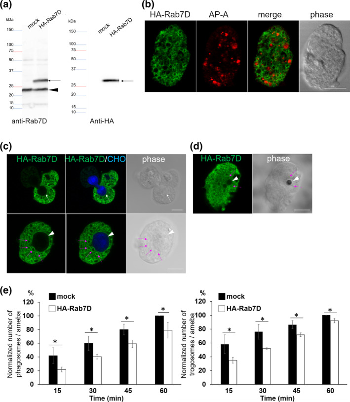
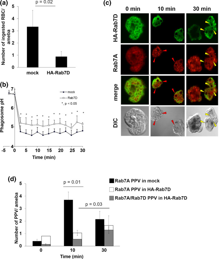
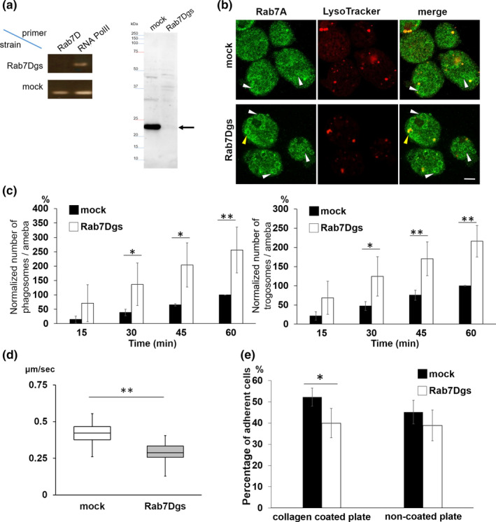
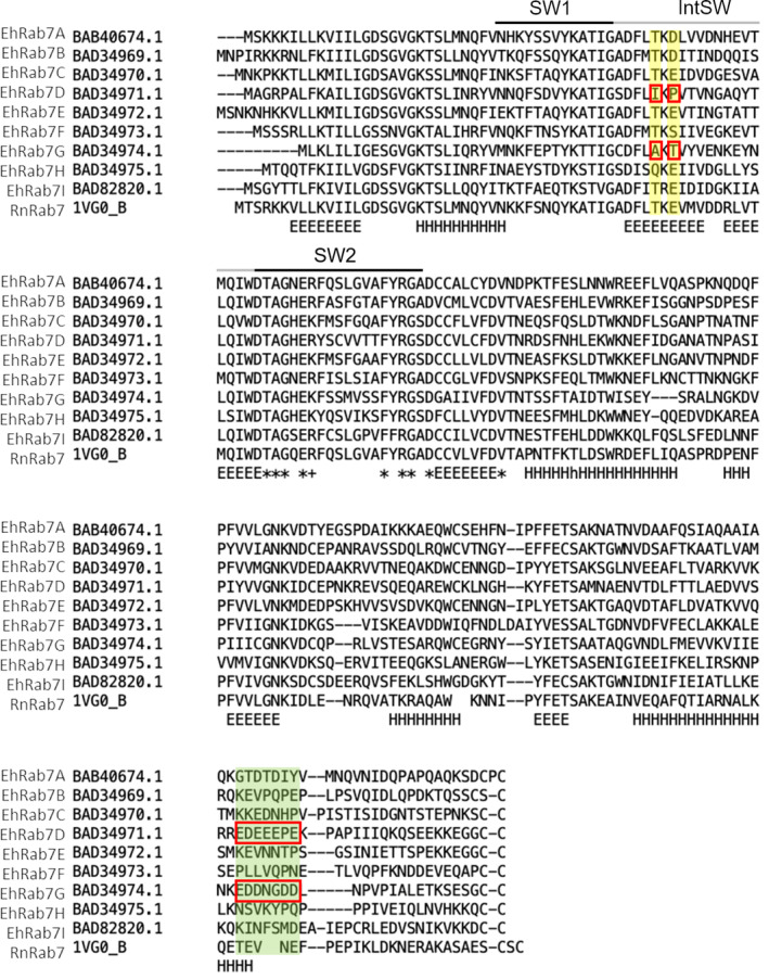
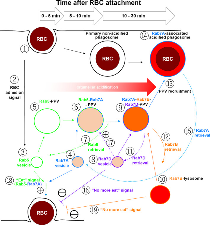
Rab small GTPases regulate membrane traffic between distinct cellular compartments of all eukaryotes in a tempo-spatially specific fashion. Rab small GTPases are also involved in the regulation of cytoskeleton and signalling. Membrane traffic and cytoskeletal regulation play pivotal role in the pathogenesis of Entamoeba histolytica, which is a protozoan parasite responsible for human amebiasis. E. histolytica is unique in that its genome encodes over 100 Rab proteins, containing multiple isotypes of conserved members (e.g., Rab7) and Entamoeba-specific subgroups (e.g., RabA, B, and X). Among them, E. histolytica Rab7 is the most diversified group consisting of nine isotypes. While it was previously demonstrated that EhRab7A and EhRab7B are involved in lysosome and phagosome biogenesis, the individual roles of other Rab7 members and their coordination remain elusive. In this study, we characterised the third member of Rab7, Rab7D, to better understand the significance of the multiplicity of Rab7 isotypes in E. histolytica. Overexpression of EhRab7D caused reduction in phagocytosis of erythrocytes, trogocytosis (meaning nibbling or chewing of a portion) of live mammalian cells, and phagosome acidification and maturation. Conversely, transcriptional gene silencing of EhRab7D gene caused opposite phenotypes in phago/trogocytosis and phagosome maturation. Furthermore, EhRab7D gene silencing caused reduction in the attachment to and the motility on the collagen-coated surface. Image analysis showed that EhRab7D was occasionally associated with lysosomes and prephagosomal vacuoles, but not with mature phagosomes and trogosomes. Finally, in silico prediction of structural organisation of EhRab7 isotypes identified unique amino acid changes on the effector binding surface of EhRab7D. Taken together, our data suggest that EhRab7D plays coordinated counteracting roles: a inhibitory role in phago/trogocytosis and lyso/phago/trogosome biogenesis, and an stimulatory role in adherence and motility, presumably via interaction with unique effectors. Finally, we propose the model in which three EhRab7 isotypes are sequentially involved in phago/trogocytosis.

Keywords: Entamoeba histolytica; Rab7D; cytoskeleton; lysosome; pathogenesis; phagocytosis; trogocytosis; vesicular traffic.


Conflict of interest statement

The authors declare no conflict of interest.

Figures

FIGURE 1
FIGURE 1
Expression and localization of HA‐EhRab7D and the effect of EhRab7D overexpression on phagocytosis and trogocytosis. (a) Immunoblot analysis of HA‐EhRab7D in E. histolytica transformants. Total cell lysates from mock‐transfected control (mock) and HA‐EhRab7D‐expressing transformant (HA‐Rab7D) were subjected to SDS‐PAGE and immunoblot analysis using the anti‐EhRab7D (left panel) and anti‐HA (right panel) antibodies. Arrows indicate HA‐EhRab7D. An arrowhead indicates intrinsic wild‐type EhRab7D. (b) Localization of HA‐EhRab7D in quiescent state. HA‐EhRab7D‐expressing transformant was fixed and reacted with anti‐HA (green) and anti‐AP‐A (red) antibodies. Bar, 10 μm. (c) Localization of HA‐EhRab7D during trogocytosis. HA‐EhRab7D expressing E. histolytica trophozoites were co‐cultured with CellTracker Blue‐stained live CHO cells for 15 min, fixed, and reacted with the anti‐HA antibody (green). A white arrow indicate trogocytic cup. An arrowhead indicate the trogosome. Magenta arrows indicate HA‐EhRab7D‐associated vesicles adjacent to the trogosome. Bar, 10 μm. (d) Localization of HA‐EhRab7D during phagocytosis of artificial beads. HA‐EhRab7D expressing E. histolytica trophozoites were incubated with human serum coated Dynabeads (2.8‐μm diameter) for 15 min, fixed, and reacted with the anti‐HA antibody (green). A white arrowhead and magenta arrows indicate the phagosome and HA‐EhRab7D‐associated vesicles, respectively. Bar, 10 μm. (e) The effect of HA‐ERab7D expression on phagocytosis and trogocytosis. Trophozoites of mock transfected and HA‐EhRab7D expressing strains were prestained with CellTracker Blue and co‐cultivated with live or heat‐killed CHO cells that had been prestained with CellTracker Orange to evaluate trogocytosis or phagocytosis, respectively. Microscopic images were captured on CQ1 every 15 min for 60 min and analysed to calculate the average numbers of CHO cell‐containing trogo‐/phagosomes per ameba. All data were normalised against the values obtained for control at 1 hr and are shown as percentages to these values. Data of one representative experiment was shown. Statistical significance was examined with Student's t test, *p < .05. n = 3
FIGURE 2
FIGURE 2
Effect of HA‐EhRab7D expression on erythrophagocytosis, phagosome acidification, and PPV formation. (a) Erythrophagocytosis by mock transfected and HA‐EhRab7D expressing strains. Trophozoites of mock transfected and HA‐EhRab7D expressing strains were incubated with hamster erythrocytes (RBCs) for 30 min. Cells were fixed and erythrocytes were visualised with diaminobenzidine. Number of RBCs per amoeba was counted. Statistical significance was inferred by t‐test, p = .02. (b) Kinetics of phagosome acidification in mock transfected (closed circles) and mRFP‐EhRab7D‐expressing (open squares) strains. Trophozoites were incubated with FITC‐conjugated yeasts at 1:10 ratio at 33°C to allow the amebae to ingest yeasts. mRFP‐EhRab7D‐expressing trophozoites that ingested only a single yeast were selected and phagosome pH was measured at 1 min intervals for 30 min. Data represent the average of 10 independent phagosomes. Statistical significance was examined with t‐test, *p < .05. (c) Localization of HA‐EhRab7D and EhRab7A during erythrophagocytosis. Trophozoites of HA‐EhRab7D‐expressing strain was incubated with RBCs for indicated time, fixed, reacted with anti‐EhRab7A or anti‐HA antibodies to visualise EhRab7A (red) or HA‐EhRab7D (green). Ingested RBCs were visualised by diaminobenzidine. Red arrowheads indicate EhRab7A‐associated vacuoles and yellow arrowheads indicate EhRab7A and HA‐EhRab7D double‐associated vacuoles. Merged images of EhRab7A and HA‐EhRab7D and differential interference contrast (DIC) images are also shown. (d) PPV formation during erythrophagocytosis in mock (black) and HA‐EhRab7D overexpressing (white and grey) transformants. Trophozoites of mock transfected and HA‐EhRab7D expressing strains were incubated with RBCs for indicated time, fixed, and reacted with anti‐EhRab7A (black and white), anti‐HA (grey), and diaminobenzidine as in (c). The numbers of EhRab7A‐associated vacuoles (PPV) and EhRab7A/HA‐RhRab7D‐double associated PPVs per ameba were calculated. Statistical significance was examined with t‐test, p = .01 and .03
FIGURE 3
FIGURE 3
Establishment and phenotypes of EhRab7D gene silenced strain. (a) Confirmation of gene silencing by reverse transcriptase (RT)‐PCR (left panel) and immunoblot (right panel) analysis of mock transfected and EhRab7D gene silenced (gs) strain. Transcripts of EhRab7D and RNA polymerase II genes were amplified by RT‐PCR from cDNA isolated from the transformants and examined by agarose gel electrophoresis (left panel). Total lysate of the transformants were subjected to SDS‐PAGE and immunoblot analysis using the anti‐EhRab7D antibody (right panel). (b) Localization of EhRab7A and LysoTracker in EhRab7D gs and control strains. EhRab7D gs and mock transfected strains were stained with LysoTracker Red, fixed, and reacted with anti‐EhRab7A antibody (green). The diameter of the largest EhRab7A‐positive vacuole in each cell was measured in 50 trophozoites from three independent experiments. (c) The effect of gene silencing of EhRab7D gene on phagocytosis (left) and trogocytosis (right). Trophozoites of mock transfected and EhRab7D gene silenced strains prestained with CellTracker Blue were co‐cultivated with heat‐killed (left panel) or live (right panel) CHO cells prestained with CellTracker Orange to evaluate phagocytosis or trogocytosis, respectively. Also see the legend in Figure 1e. Statistical significance was examined with t‐test, *p < .05, **p < .01. n = 4. (d) Cell motility of mock transfected and EhRab7D gene silenced strain. The trophozoites of the indicated strains were stained with CellTracker Green were incubated with BI medium at 35.5°C on a collagen‐coated glass‐bottom dish. Time‐lapse images were collected every 30 s for 15 min on LSM780 and processed using Imaris software. Data form one representative experiment was shown. Statistical significance was examined with Dunnet test, **p < .05. n = 2. (e) Adhesion to collagen coated and non‐coated plastic plates of mock transfected and EhRab7D gene silenced strain. Trophozoites were prestained with CellTracker Green and incubated with BI medium on indicated plates at 35.5°C for 40 min. After incubation, the plates were gently rinsed with fresh warm BI medium, and the fluorescence intensity of each well before and after washing was measured, normalised against the value of the mock control at 60 min, and is shown in percentage. Statistical significance was examined with t‐test, *p < .05 and n = 4
FIGURE 4
FIGURE 4
Alignment of nine EhRab7 isotypes and rat Rab7 (RnRab7). Multiple amino Acid sequence alignment of all nine E. histolytica Rab7 isotypes with rat Rab7 (RnRab7, PDB ID: 1vg0B/UniProt: P09527) was constructed by using FAMSA with its default parameters. Switch 1 (SW1), switch 2 (SW2), and inter switch (IntSW) regions are depicted above the sequences. Two regions containing EhRab7D‐ and EhRab7G‐specific amino acid substitutions are coloured in yellow or green, and unique amino acids are highlighted with red squares. A region with yellow background corresponds to the interface between mammalian Rab7 effector RILP (see Supplementary Figure S2). Note the conserved motif of T/Q‐K/R‐E/D/S in all sequences except for EhRab7D and EhRab7G, in which the motif is replaced with I‐K‐P or A‐K‐T, respectively. The region with green background is located in an intrinsically disordered region, in which EhRab7D and EhRab7G contain a unique acidic amino acid stretch. “E” and “H” denote beta‐strand and alpha‐helix, respectively. “*” and “+” depict completely conserved and positively charged amino acids around SW2
FIGURE 5
FIGURE 5
A proposed model of the roles of EhRab7A, EhRab7B, and EhRab7D during erythrophagocytosis of E. histolytica. Refer to Discussion

Similar articles

Cited by

References

    1. Agarwal, S. , Anand, G. , Sharma, S. , Parimita Rath, P. , Gourinath, S. , & Bhattacharya, A. (2019). EhP3, a homolog of 14‐3‐3 family of protein participates in actin reorganization and phagocytosis in Entamoeba histolytica . PLoS Pathogens, 15(5), e1007789 10.1371/journal.ppat.1007789 - DOI - PMC - PubMed
    1. Aguilar‐Rojas, A. , Olivo‐Marin, J. C. , & Guillen, N. (2016). The motility of Entamoeba histolytica: Finding ways to understand intestinal amoebiasis. Current Opinion in Microbiology, 34, 24–30. 10.1016/j.mib.2016.07.016 - DOI - PubMed
    1. Babuta, M. , Bhattacharya, S. , & Bhattacharya, A. (2020). Entamoeba histolytica and pathogenesis: A calcium connection. PLoS Pathogens, 16(5), e1008214 10.1371/journal.ppat.1008214 - DOI - PMC - PubMed
    1. Babuta, M. , Kumar, S. , Gourinath, S. , Bhattacharya, S. , & Bhattacharya, A. (2018). Calcium‐binding protein EhCaBP3 is recruited to the phagocytic complex of Entamoeba histolytica by interacting with Arp2/3 complex subunit 2. Cellular Microbiology, 20(12), e12942 10.1111/cmi.12942 - DOI - PubMed
    1. Babuta, M. , Mansuri, M. S. , Bhattacharya, S. , & Bhattacharya, A. (2015). The Entamoeba histolytica, Arp2/3 complex is recruited to phagocytic cups through an atypical kinase EhAK1. PLoS Pathogens, 11(12), e1005310 10.1371/journal.ppat.1005310 - DOI - PMC - PubMed

Publication types

MeSH terms

Substances