. 2020 Oct;48(7):887-898.
doi: 10.1177/0192623320961017. Epub 2020 Sep 25.

Ozone Reacts With Carbon Black to Produce a Fulvic Acid-Like Substance and Increase an Inflammatory Effect

Affiliations

Ozone Reacts With Carbon Black to Produce a Fulvic Acid-Like Substance and Increase an Inflammatory Effect

Andrew J Ghio et al. Toxicol Pathol. 2020 Oct.

Abstract

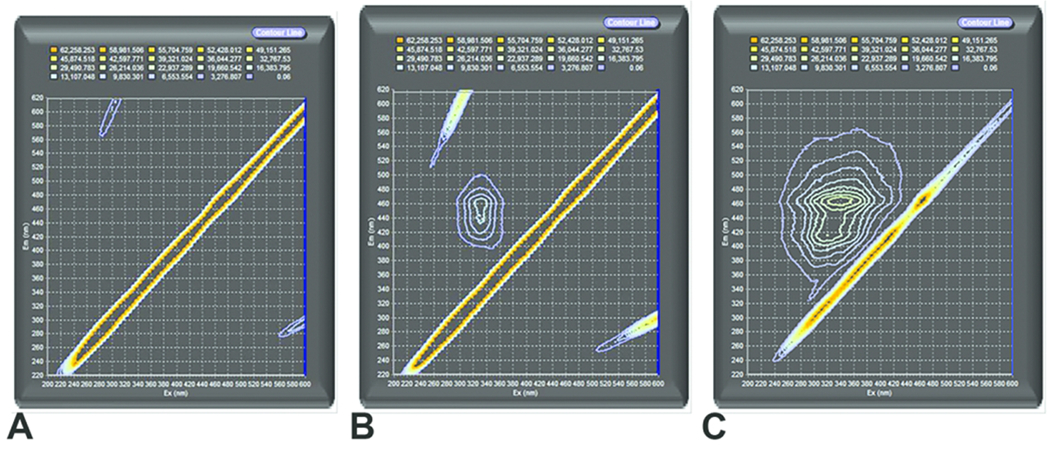
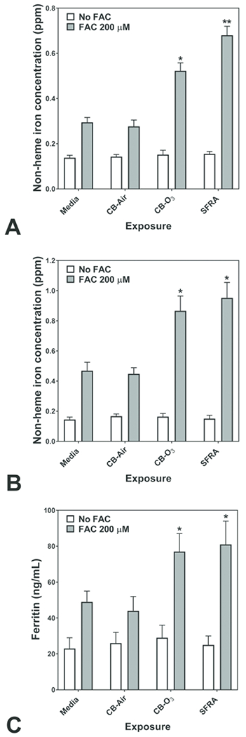
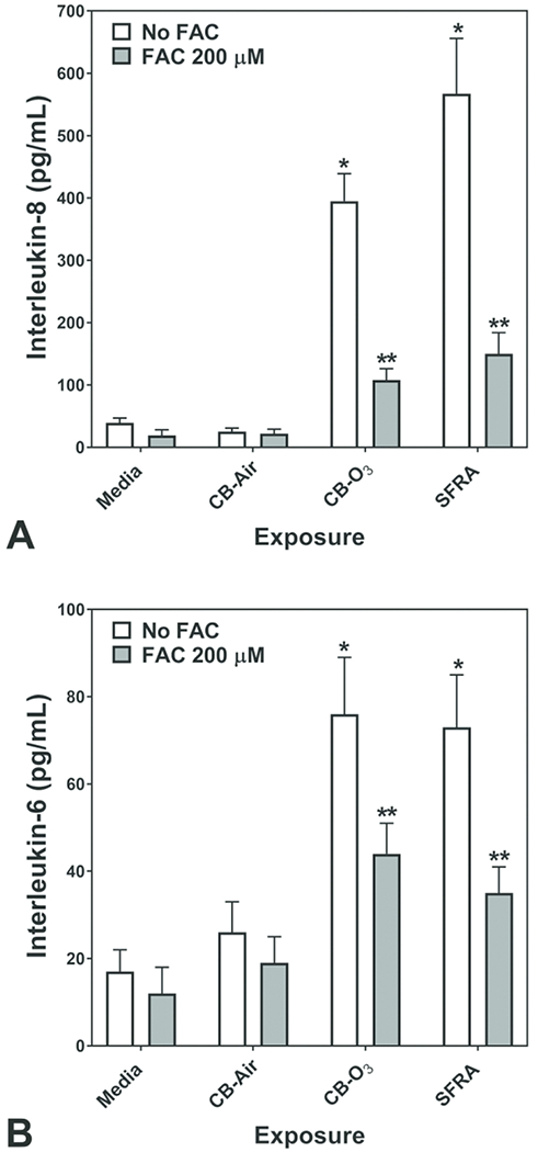
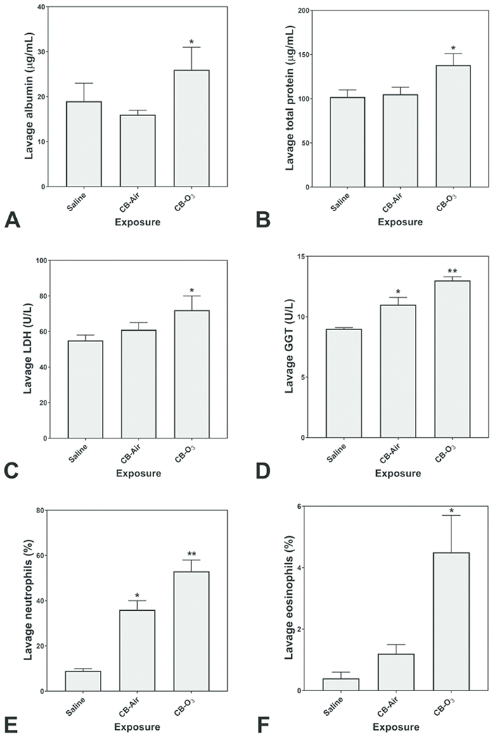
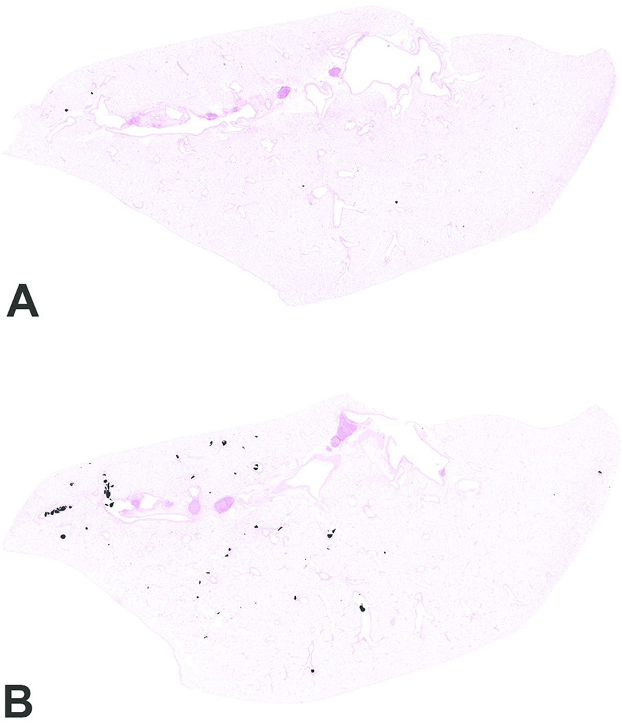
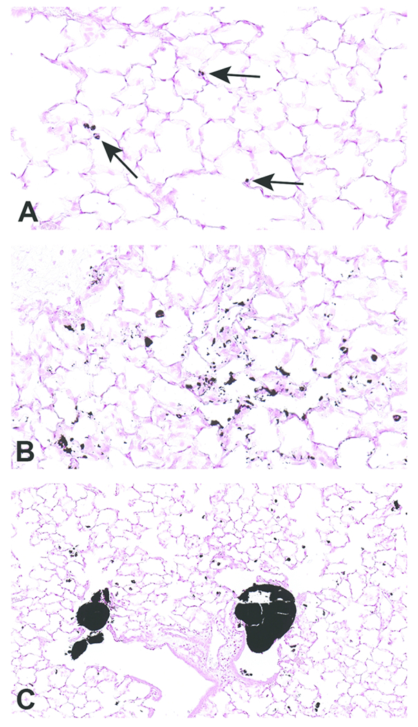
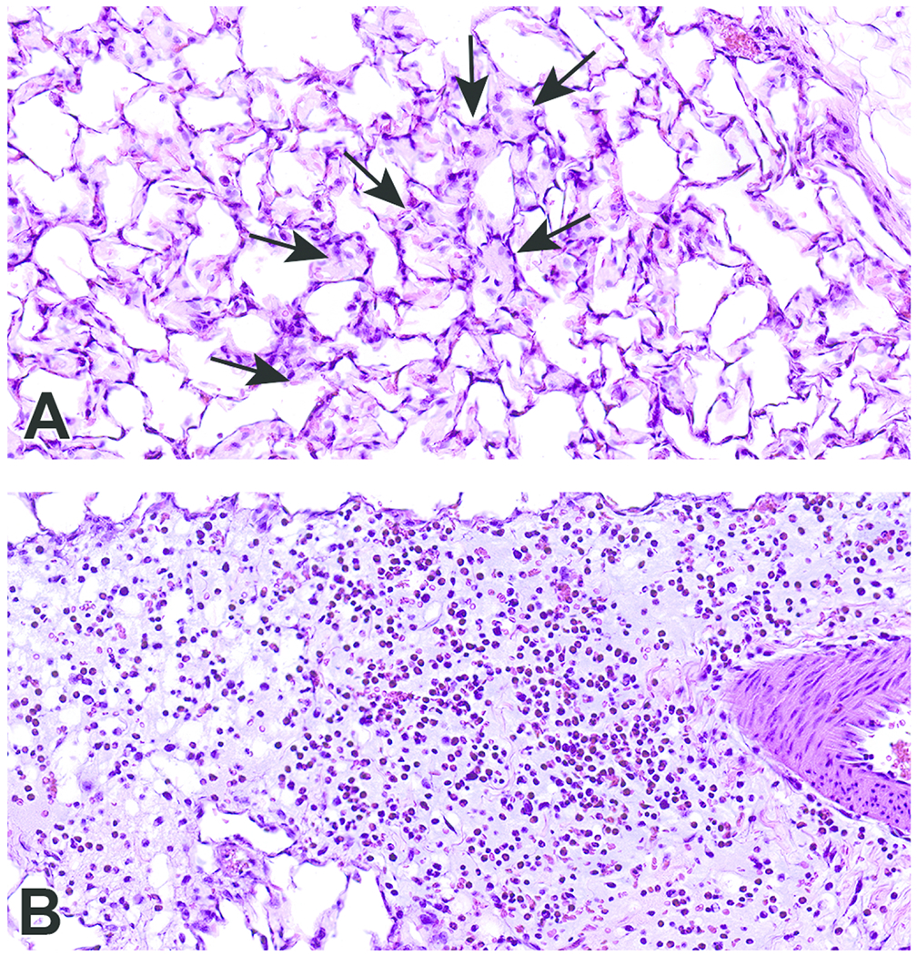
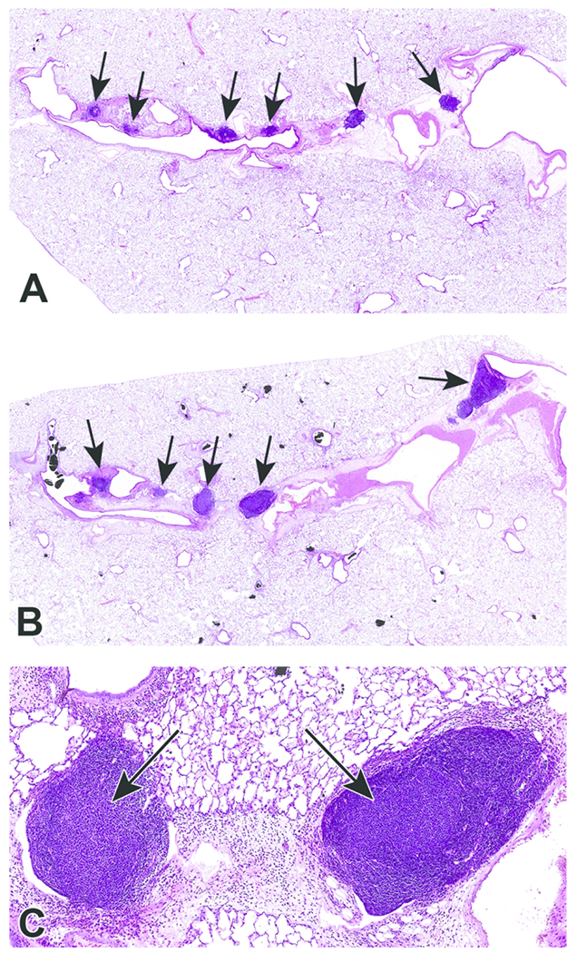
Exposure to ambient ozone has been associated with increased human mortality. Ozone exposure can introduce oxygen-containing functional groups in particulate matter (PM) effecting a greater capacity of the particle for metal complexation and inflammatory effect. We tested the postulate that (1) a fulvic acid-like substance can be produced through a reaction of a carbonaceous particle with high concentrations of ozone and (2) such a fulvic acid-like substance included in the PM can initiate inflammatory effects following exposure of respiratory epithelial (BEAS-2B) cells and an animal model (male Wistar Kyoto rats). Carbon black (CB) was exposed for 72 hours to either filtered air (CB-Air) or approximately 100 ppm ozone (CB-O3). Carbon black exposure to high levels of ozone produced water-soluble, fluorescent organic material. Iron import by BEAS-2B cells at 4 and 24 hours was not induced by incubations with CB-Air but was increased following coexposures of CB-O3 with ferric ammonium citrate. In contrast to CB-Air, exposure of BEAS-2B cells and rats to CB-O3 for 24 hours increased expression of pro-inflammatory cytokines and lung injury, respectively. It is concluded that inflammatory effects of carbonaceous particles on cells can potentially result from (1) an inclusion of a fulvic acid-like substance after reaction with ozone and (2) changes in iron homeostasis following such exposure.

Keywords: air pollution; carbon black; fulvic acid; fulvic acid-like substance; inflammation; iron; lung diseases; ozone; rats.


Conflict of interest statement

Declaration of conflicting interest statement

The authors declare no potential conflicts of interest with respect to the research, authorship, and/or publication of this article.

Figures

Figure 1.
Figure 1.
Fluorescence excitation-emission matrix spectra of carbon black treated with air (CB-Air) (A), carbon black treated with ozone (CB-O3) (B), and Suwanee River fulvic acid (SRFA) (C). The exposure of CB to high levels of ozone produced water-soluble, fluorescent organic material with similarities of fluorophores with SRFA. The diagonal line towards the right lower corner is an instrument artifact due to light scattering.
Figure 2.
Figure 2.
Cell non-heme iron concentrations following 4 (A) and 24 (B) hr exposures of BEAS-2B cells to media, carbon black treated with air (CB-Air), carbon black treated with ozone (CB-O3), and Suwanee River fulvic acid (SRFA). Incubations were without and with 200 μM FAC. Exposures to CB-Air, CB-O3, and SRFA did not impact cell non-heme iron relative to media at either time point. Exposure to FAC increased cell non-heme iron but co-exposure with CB-O3, was associated with significantly increased non-heme iron concentrations at both 4 (A) and 24 (B) hr. Exposures of BEAS-2B cells to FAC increased cell ferritin levels after 24 hr relative to media alone (C). However, co-exposure of CB-O3, and SRFA with FAC was associated with significantly elevated cell ferritin concentrations (C). Two-way ANOVA indicated that both addition of FAC and exposure group significantly impacted cell non-heme iron. Among the cell exposures with addition of FAC, one-way ANOVA demonstrated: * significant increase relative to exposure of BEAS-2B cells to media only and ** significant increase relative to all other exposures.
Figure 3.
Figure 3.
Release of IL-8 and IL-6 after 24 hr exposure of BEAS-2B cells to media, carbon black treated with air (CB-Air), carbon black treated with ozone (CB-O3), and Suwanee River fulvic acid (SRFA). Exposure to CB-O3, and SRFA significantly increased the release of IL-8 and IL-6. FAC treatment significantly diminished this response. One-way ANOVA demonstrated: * significant increase relative to all other exposures and ** significant increase relative to media exposure.
Figure 4.
Figure 4.
Lavage indices of injury and inflammation following animal exposures to normal saline, carbon black treated with air (CB-Air), and carbon black treated with ozone (CB-O3). There is evidence of both injury and inflammation following both CB-Air and CB-O3 but both are significantly greater following CB-O3. One-way ANOVA demonstrated: * significant increase relative to exposure of animals to saline only and ** significant increase relative to all other exposures.
Figure 5.
Figure 5.
Scanned images of lung pathology following animal exposures to carbon black treated with air (CB-Air; A) or ozone (CB-O3; B). There are increased numbers of particles and the particles are larger in size in CB-O3 (B) animals compared to CB-Air (A) animals. Tissues were stained with Prussian blue to better illustrate the particles. Original scans at 1x.
Figure 6.
Figure 6.
Scanned images of lung pathology following exposures to carbon black treated with air (CB-Air; A) or ozone (CB-O3; B). Figures 6A and 6B are higher magnification images (original scans at 40x) of figures 5A and 5B, respectively. Particles in A are indicated with arrows. There was an increased number and size of particles in CB-O3 (B) lungs compared to CB-Air (A) lungs. Figure C illustrates the scattered larger particles in CB-O3 lungs (original scan at 20x). The larger aggregates were predominately in the terminal bronchi and alveolar ducts. Tissues were stained with Prussian blue to better illustrate the particles.
Figure 7.
Figure 7.
Scanned images show examples of macrophage infiltrates within alveoli (A; arrows) and perivascular and peribronchiolar interstitial inflammation (B) in animals exposed to CB-O3. Edema is present in the perivascular and peribronchiolar regions of inflammation. Macrophages and granulocytic infiltrates were increased in the CB-O3 group compared to the CB-air group. Original scans at 40x; stain is hematoxylin and eosin.
Figure 8.
Figure 8.
Photomicrographs of bronchioalveolar lymphoid aggregates (BALT) in animals exposed to CB-Air (A) and CB-O3 (B). BALT was increased in the CB-O3 (B; arrows) group relative to the CB-Air group (A; arrows) and CB-O3 lungs typically had prominent germinal centers (C; arrows). A and B original scans at 2x while C original scan at 14x; stain is hematoxylin and eosin.

Similar articles

Cited by

References

    1. Ghio AJ, Tong H, Soukup JM, et al. Sequestration of mitochondrial iron by silica particle initiates a biological effect. Am J Physiol Lung Cell Mol Physiol. 2013;305(10):L712–724. - PubMed
    1. Ghio AJ, Soukup JM, Dailey LA, Madden MC. Air pollutants disrupt iron homeostasis to impact oxidant generation, biological effects, and tissue injury. Free Radic Biol Med. 2020;151:38–55. - PMC - PubMed
    1. Laughton MJ, Moroney MA, Hoult JR, Halliwell B. Effects of desferrioxamine on eicosanoid production in two intact cell systems. Biochem Pharmacol. 1989;38(1):189–193. - PubMed
    1. Hileti D, Panayiotidis P, Hoffbrand AV. Iron chelators induce apoptosis in proliferating cells. Br J Haematol. 1995;89(1):181–187. - PubMed
    1. Tanji K, Imaizumi T, Matsumiya T, et al. Desferrioxamine, an iron chelator, upregulates cyclooxygenase-2 expression and prostaglandin production in a human macrophage cell line. Biochim Biophys Acta. 2001;1530(2–3):227–235. - PubMed

Publication types