. 2020 Oct;6(10):mgen000442.
doi: 10.1099/mgen.0.000442.

Expanded roles of pyruvate-sensing PdhR in transcription regulation of the Escherichia coli K-12 genome: fatty acid catabolism and cell motility

Affiliations

Expanded roles of pyruvate-sensing PdhR in transcription regulation of the Escherichia coli K-12 genome: fatty acid catabolism and cell motility

Takumi Anzai et al. Microb Genom. 2020 Oct.

Abstract

The transcription factor PdhR has been recognized as the master regulator of the pyruvate catabolism pathway in Escherichia coli, including both NAD-linked oxidative decarboxylation of pyruvate to acetyl-CoA by PDHc (pyruvate dehydrogenase complex) and respiratory electron transport of NADH to oxygen by Ndh-CyoABCD enzymes. To identify the whole set of regulatory targets under the control of pyruvate-sensing PdhR, we performed genomic SELEX (gSELEX) screening in vitro. A total of 35 PdhR-binding sites were identified along the E. coli K-12 genome, including previously identified targets. Possible involvement of PdhR in regulation of the newly identified target genes was analysed in detail by gel shift assay, RT-qPCR and Northern blot analysis. The results indicated the participation of PdhR in positive regulation of fatty acid degradation genes and negative regulation of cell mobility genes. In fact, GC analysis indicated an increase in free fatty acids in the mutant lacking PdhR. We propose that PdhR is a bifunctional global regulator for control of a total of 16-23 targets, including not only the genes involved in central carbon metabolism but also some genes for the surrounding pyruvate-sensing cellular pathways such as fatty acid degradation and flagella formation. The activity of PdhR is controlled by pyruvate, the key node between a wide variety of metabolic pathways, including generation of metabolic energy and cell building blocks.

Keywords: Escherichia coli; PdhR; cell motility; fatty acid β-oxidation; gSELEX; pyruvate; transcription regulation.


Conflict of interest statement

The authors declare that there are no conflicts of interest.

Figures
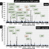
Fig. 1.
Fig. 1.
Identification of PdhR-binding sites on the E. coli K-12 genome by gSELEX-chip analysis. Genomic SELEX screening of PdhR-binding sequences was performed in the absence (a) or presence (b) of pyruvate. After gSELEX, a collection of DNA fragments was subjected to gSELEX-chip analysis using the tiling array of the E. coli K-12 genome. The y-axis represents the relative number of PdhR-bound DNA fragments, and the x-axis represents the position on the E. coli genome. The regulation target genes of PdhR at high-level peaks are shown along the E. coli genome. Peaks shown in green represent the type-A and type-B PdhR-binding sites inside spacer regions, while peaks shown in orange represent the type-D PdhR-binding sites inside ORFs (for typing see the legend to Table 1). The cut-off level of 500 is shown by a blue line, and the list of all PdhR-binding sites by setting this cut-off level is given in Table 1.
Fig. 2.
Fig. 2.
Confirmation of the PdhR-binding in vitro to the regulatory targets: gel shift assay. Purified His-tagged PdhR was mixed with 0.2 pmol of each DNA probe corresponding to the PdhR-binding regions shown in Fig. 1. PdhR (pmol) was added: lane 1, 0; lane 2, 1.25; lane 3, 2.5; lane 4, 5.0; lane 5, 10.0. Filled triangles indicate the PdhR–DNA probe complex while open triangles indicate free probe.
Fig. 3.
Fig. 3.
Consensus sequence of the PdhR-box. Sequences of all the probes with high-level binding of PdhR were analysed using dminda 2.0 (http://bmbl.sdstate.edu/DMINDA2/) (see Table 1). (a) Sequences built with the PdhR-target sequences listed in RegulonDB. (b) weblogo (http:// weblogo.berkeley.edu/logo.cgi) was used for matrix construction.
Fig. 4.
Fig. 4.
Influence of pdhR deletion on transcription of the newly identified PdhR targets by RT-qPCR. E. coli wild-type BW25113 and the pdhR-deleted mutant JW0109 were grown in M9 medium with 0.2 % succinate. Total RNA was prepared from both the wild-type and pdhR mutant, and subjected to RT-qPCR analysis (see Experimental procedures). RT-qPCR was repeated three times and the average means are shown. This list shows mRNA levels of the indicated regulation target genes of PdhR. The y-axis represents the relative level of mRNA of each PdhR target gene between wild-type and the pdhR mutant, by setting the ratio of 16S rRNA as an internal control between wild-type and the phdR mutant.
Fig. 5.
Fig. 5.
Motility assay on soft agar plates. Transformants of E. coli wild-type BW25113, pdhR-deleted mutant JW0109 with PdhR expression plasmid or its control empty vector pCA24NΔgfp were inoculated on the 0.25 % agar plate in M9 minimal medium with 0.2 % succinate in the absence (a1) or presence (a2) of 50 µM IPTG. After 24 h of incubation, the plate image was observed (a) and the colony diameter was measured as the migration zone (b).
Fig. 6.
Fig. 6.
Amount of free fatty acids: individual fatty acids and total fatty acids. E. coli wild-type BW25113, pdhR-deleted mutant JW0109 and fadR-deleted mutant JW1176 were grown in M9 minimal medium with 0.2 % succinate. Total free fatty acids were extracted from exponentially growing cells, and purified by TLC. The amount of each fatty acid was analysed with GC-FID (a). The total amount of free fatty acids was calculated as the sum of individual fatty acids (b). The y-axis (μg l−1/OD600) represents the amount of free fatty acid. The levels of fatty acids in three E. coli strains are shown separately: black, wild-type; white, pdhR mutant; grey, fadR mutant.
Fig. 7.
Fig. 7.
Influence of pdhR deletion and pyruvate addition on transcription of the newly identified PdhR targets for fatty acid degradation. E. coli wild-type BW25113 and the pdhR-deleted mutant JW0109 were grown in M9 minimal medium with 0.2 % succinate in the absence and presence of 3 mM pyruvate. The relative level of mRNA of each PdhR target gene between wild-type and pdhR mutant is shown by black bars, while the relative level of mRNA in wild-type E. coli in the presence and absence of pyruvate is shown by white bars. The experiment was repeated three times and the average values are shown.
Fig. 8.
Fig. 8.
Northern blot analysis of mRNAs for fatty acid degradation. E. coli wild-type BW25113 and its pdhR mutant JW0109 were grown in M9 minimal medium with 0.2 % succinate. Total RNA was prepared at the exponential phase and directly subjected to Northern blot analysis under the standard conditions as described in the Methods. DIG-labelled hybridization probes are shown on the left side of each panel. The amounts of PdhR target mRNAs were measured as relative levels of 23S and 16S rRNAs stained with methylene blue.
Fig. 9.
Fig. 9.
Model of the PdhR regulon. The whole set of regulatory targets of PdhR obtained in this study are aligned along the E. coli metabolism pathway. The PdhR targets include the genes involved in carbon source catabolism from PDH down to respiratory electron transport, lactate degradation, TCA cycle and β-oxidation of fatty acid degradation. The genes repressed by PdhR are shown in blue while the genes activated by PdhR are shown in red. The PdhR regulon members, ppsA, pykF, lldP, lldD, fumD, fadE, fadI and fadJ, were identified in this study.

References

    1. Haydon DJ, Quail MA, Guest JR. A mutation causing constitutive synthesis of the pyruvate dehydrogenase complex in Escherichia coli is located within the pdhR gene. FEBS Lett. 1993;336:43–47. doi: 10.1016/0014-5793(93)81605-Y. - DOI - PubMed
    1. Quail MA, Guest JR. Purification, characterization and mode of action of PdhR, the transcriptional repressor of the pdhR-aceEF-lpd operon of Escherichia coli . Mol Microbiol. 1995;15:519–529. doi: 10.1111/j.1365-2958.1995.tb02265.x. - DOI - PubMed
    1. Ogasawara H, Ishida Y, Yamada K, Yamamoto K, Ishihama A. PdhR (pyruvate dehydrogenase complex regulator) controls the respiratory electron transport system in Escherichia coli . J Bacteriol. 2007;189:5534–5541. doi: 10.1128/JB.00229-07. - DOI - PMC - PubMed
    1. Göhler AK, Kökpinar O, Schmidt-Heck W, Geffers R, Guthke R, et al. More than just a metabolic regulator--elucidation and validation of new targets of PdhR in Escherichia coli . BMC Syst Biol. 2011;5:197. doi: 10.1186/1752-0509-5-197. - DOI - PMC - PubMed
    1. Salgado H, Gama-Castro S, Martínez-Antonio A, Díaz-Peredo E, Sánchez-Solano F, et al. RegulonDB (version 4.0): transcriptional regulation, operon organization and growth conditions in Escherichia coli K-12. Nucleic Acids Res. 2004;32:303D–306. doi: 10.1093/nar/gkh140. - DOI - PMC - PubMed

Publication types

MeSH terms

LinkOut - more resources