. 2021 Feb 2;72(2):184-198.
doi: 10.1093/jxb/eraa444.

Blocking intruders: inducible physico-chemical barriers against plant vascular wilt pathogens

Affiliations

Blocking intruders: inducible physico-chemical barriers against plant vascular wilt pathogens

Anurag Kashyap et al. J Exp Bot. .

Abstract

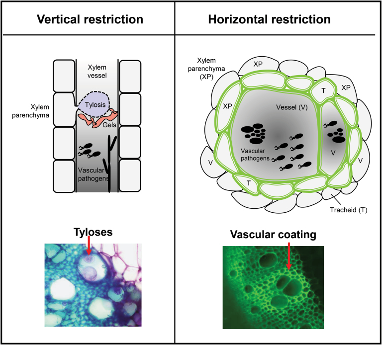
Xylem vascular wilt pathogens cause devastating diseases in plants. Proliferation of these pathogens in the xylem causes massive disruption of water and mineral transport, resulting in severe wilting and death of the infected plants. Upon reaching the xylem vascular tissue, these pathogens multiply profusely, spreading vertically within the xylem sap, and horizontally between vessels and to the surrounding tissues. Plant resistance to these pathogens is very complex. One of the most effective defense responses in resistant plants is the formation of physico-chemical barriers in the xylem tissue. Vertical spread within the vessel lumen is restricted by structural barriers, namely, tyloses and gels. Horizontal spread to the apoplast and surrounding healthy vessels and tissues is prevented by vascular coating of the colonized vessels with lignin and suberin. Both vertical and horizontal barriers compartmentalize the pathogen at the infection site and contribute to their elimination. Induction of these defenses are tightly coordinated, both temporally and spatially, to avoid detrimental consequences such as cavitation and embolism. We discuss current knowledge on mechanisms underlying plant-inducible structural barriers against major xylem-colonizing pathogens. This knowledge may be applied to engineer metabolic pathways of vascular coating compounds in specific cells, to produce plants resistant towards xylem colonizers.

Keywords: Gels; inducible defenses; lignin; physico-chemical barriers; plant-pathogen interactions; structural defenses; suberin; tyloses; vascular pathogens; wilt.


Figures

Fig. 1.
Fig. 1.
The two dimensions of plant physico-chemical barriers induced against xylem vascular wilt pathogens. To counter invasion by xylem vascular wilt pathogens, resistant plants induce two-dimensional physico-chemical defenses that restrict vertical and horizontal movement of the pathogen. Vertical spread within the vessel lumen is mainly restricted by tyloses and gels (left). In contrast, horizontal spread of the pathogen to surrounding healthy vessels is prevented by reinforcement of the walls of colonized vessels (V) and the surrounding xylem parenchyma (XP) and tracheids (T), through vascular coating with mainly lignin and suberin (shown as a green color in the diagram). Synchronized formation of vertical and horizontal barriers early after pathogen invasion results in compartmentalization of the pathogen inoculum at the site of infection, thereby preventing wilt, and constitute a major component of resistance. To visualize tyloses and vascular coating (panels below), tomato root cross-sections were obtained after R. solanacearum soil soak inoculation, and fixed in 70% ethanol. For tyloses, cross-sections were stained with 0.1% toluidine blue and observed using a Leica DM6B-Z microscope under bright field conditions, and images were recorded through a MC190-HD-0518131623 camera. To visualize phenolic vascular coating, the cross-sections were illuminated by UV using a Leica DM6B-Z microscope, and the auto-florescence emitted from phenolic deposits was observed using a HC PL FLUOTAR objective. Images were captured using a Leica-DFC9000GT-VSC07341 camera. In the left panel, the arrow points towards R. solanacearum-induced formation of tyloses inside vessel lumen, which appear pink to violet color upon staining with 0.1% toluidine blue. In the right panel, the arrow points towards R. solanacearum-induced auto-fluorescence emitted from phenolics, deposited in the walls of vessels and the surrounding tracheids and parenchyma cells. Scale bar=120 µm.
Fig. 2.
Fig. 2.
Lignin and suberin have a major role in vascular coating induced by xylem vascular wilt pathogens. A. Schematic structure of reinforced cell walls of xylem vessels and parenchyma cells in resistant plants upon infection with xylem vascular wilt pathogens. B. The phenypropanoid pathway provides precursors for both lignin and suberin biosynthesis. Phenylalanine, derived from the shikimate pathway, undergoes several enzymatic reactions as part of the phenylproanoid pathway. The resulting precursors yield the monolignols p-coumaryl alcohol, coniferyl alcohol and sinapyl alcohol, which are the building blocks of lignin. In parallel, the phenypropanoid metabolites feruloyl-CoA, caffeoyl-CoA, and p-coumaroyl-CoA bifurcate into the suberin pathway. In the suberin pathway, these metabolites can be conjugated to aromatic amine compounds such as tyramine by the action of THT, or can be linked to aliphatic compounds by the action of FHT, to yield suberin monomers. Lignin and suberin monomers are then transported to the cell wall, where they are subsequently polymerized into the reinforcing matrices that constitute vascular coating structures. Abbreviations: PAL: phenylalanine ammonia–lyase; C4H: cinnamate–4–hydroxylase; C3H: coumarate 3-hydroxylase; 4CL: 4–coumarate–CoA ligase; HCT: hydroxycinnamoyl–CoA shikimate/quinate hydroxycinnamoyl transferase; COMT: caffeic acid 3-O-methyltransferase; CCOMT: caffeoyl CoA 3-O-methyltransferase; CCR: cinnamoyl CoA reductase; CAD: cinnamoyl alcohol dehydrogenase; PRX: peroxidase; CYP86A1: fatty acid cytochrome P450 oxidases; FAR: fatty acyl-CoA reductase; TyDC: tyrosine decarboxylase; FHT: feruloyl transferase; THT: tyramine hydroxycinnamoyl transferase.

Similar articles

Cited by

References

    1. Alassimone J, Fujita S, Doblas VG, et al. . 2016. Polarly localized kinase SGN1 is required for Casparian strip integrity and positioning. Nature Plants 2, 16113. - PubMed
    1. Álvarez B, Biosca EG, López MM. 2010. On the life of Ralstonia solanacearum, a destructive bacterial plant pathogen. In: Méndez-Vilas A, ed. Technology and education topics in applied microbiology and microbial biotechnology. Current Research, Technology and Education Topics in Applied Microbiology. Badajoz: Formatex, 267–279.
    1. Antanaviciute L, Šurbanovski N, Harrison N, McLeary KJ, Simpson DW, Wilson F, Sargent DJ, Harrison RJ. 2015. Mapping QTL associated with Verticillium dahliae resistance in the cultivated strawberry (Fragaria × ananassa). Horticulture Research 2, 15009. - PMC - PubMed
    1. Araujo L, Bispo WM, Cacique IS, Moreira WR, Rodrigues FÁ. 2014. Resistance in mango against infection by Ceratocystis fimbriata. Phytopathology 104, 820–833. - PubMed
    1. Baayen RP, Elgersma DM. 1985. Colonization and histopathology of susceptible and resistant carnation cultivars infected with Fusarium oxysporum f. sp. dianthi. Netherlands Journal of Plant Pathology 91, 119–135.

Publication types