. 2020 Nov 4;48(19):e110.
doi: 10.1093/nar/gkaa769.

HydraPsiSeq: a method for systematic and quantitative mapping of pseudouridines in RNA

Affiliations

HydraPsiSeq: a method for systematic and quantitative mapping of pseudouridines in RNA

Virginie Marchand et al. Nucleic Acids Res. .

Abstract

Developing methods for accurate detection of RNA modifications remains a major challenge in epitranscriptomics. Next-generation sequencing-based mapping approaches have recently emerged but, often, they are not quantitative and lack specificity. Pseudouridine (ψ), produced by uridine isomerization, is one of the most abundant RNA modification. ψ mapping classically involves derivatization with soluble carbodiimide (CMCT), which is prone to variation making this approach only semi-quantitative. Here, we developed 'HydraPsiSeq', a novel quantitative ψ mapping technique relying on specific protection from hydrazine/aniline cleavage. HydraPsiSeq is quantitative because the obtained signal directly reflects pseudouridine level. Furthermore, normalization to natural unmodified RNA and/or to synthetic in vitro transcripts allows absolute measurements of modification levels. HydraPsiSeq requires minute amounts of RNA (as low as 10-50 ng), making it compatible with high-throughput profiling of diverse biological and clinical samples. Exploring the potential of HydraPsiSeq, we profiled human rRNAs, revealing strong variations in pseudouridylation levels at ∼20-25 positions out of total 104 sites. We also observed the dynamics of rRNA pseudouridylation throughout chondrogenic differentiation of human bone marrow stem cells. In conclusion, HydraPsiSeq is a robust approach for the systematic mapping and accurate quantification of pseudouridines in RNAs with applications in disease, aging, development, differentiation and/or stress response.


Figures

Figure 1.
Figure 1.
HydraPsiSeq protocol for ψ mapping and quantification. (A) General overview of the protocol. RNA is treated by hydrazine and subjected to aniline cleavage. 3′-phosphates are removed by T4 PNK treatment and adapters are ligated to 3′- and 5′-ends of RNA fragments. After sequencing and alignment to a reference sequence, 5′-ends of all fragments are counted to generate Ucleavage profiles. U residues are sensitive to hydrazine and thus efficiently cleaved, while ψ residues are resistant and provide only background signals. (B) Normalized U count for all 4 RNA nucleotides (A, C, G, U). Middle and right panels show NormUcount values for A/C/G nucleotides (N), U residues and ψ residues, in WT and strain expressing Cbf5 catalytic mutant (no Cbf5 activity, rRNA is not pseudouridylated). (C and D) Cleavage profiles for position ψ106/ψ120 in 18S rRNA in WT yeast strain and in the strain expressing Cbf5 catalytic mutant. Modified positions are shown in red and orange. U-only profiles are shown in inserts.
Figure 2.
Figure 2.
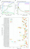
Pseudouridine detection in WT yeast rRNA. (A) Receiver Operating Characteristics (ROC) curve for ψ detection using U protection score (dark blue). Maximal Matthews correlation coefficient (MCCmax) value is shown (light blue). The corresponding trace for unmodified rRNA from Cbf5 catalytic mutant is shown in dark green (MCC trace in light green). (B) ROC curves for ψ detection for samples with variable total RNA input. As low as 50 ng of input RNA (∼40 ng of rRNA) is sufficient for good representativity and quantification of all rRNA ψ sites. (C) Dispersion of PsiScore values for technical and biological replicates. Analysis of S. cerevisiae rRNA pseudouridylation was done in three biological replicates (indicated in colors) with color bar representing dispersion of technical replicates.
Figure 3.
Figure 3.
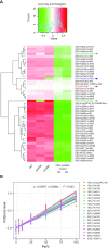
(A) Differential heatmap for variations of PsiScore level for all known sited in S. cerevisiae rRNA. Normalization was done by the average score for a given position. SnoRNA independent ψ50 in 5S rRNA is highlighted in red. ψ106 and ψ1056 guided by snR44 are highlighted in green. The identity of the S. cerevisiae strains used is provided at the bottom. Strain expressing Cbf5 catalytic mutant was analyzed in biological replicate. Identity of H/ACA snoRNA potentially guiding pseudouridylation is indicated together with the position. Clustering was done by hclust R function with ward.D2 algorithm. Color key for differential PsiScore is given in the insert. Visible variation of the signal for ψ1187 is very minor upon CBF5 inactivation, but is related to drastic change of protection at the neighboring m1acp3ψ1191, and thus perturbation in the shape of Uprofile, used to calculate PsiScore. (B) – Linear correlation of PsiScore with ψ content of yeast rRNA. Calibration curve was produced using mixes of Cbf5cata and WT rRNAs, at ratio of 0, 5, 10, 25, 50, 75 and 100% of WT rRNA. HydraPsiSeq analysis was done in technical replicate, calibration curves for 19 non-clustered ψ sites are shown.
Figure 4.
Figure 4.
Profiling of yeast mRNA and human rRNA pseudouridylation by HydraPsiSeq. (A) Profiling of yeast S. cerevisiae mRNA modifications by HydraPsiSeq. Protection profiles with sufficient coverage were obtained for 67 positions previously reported to be pseudouridylated (31,38). Out of those, 23 showing moderate to high protection (UScores from 0.5–10) are common with the list from Carlile et al (2014) and 16 with Schwartz et al. (2014), two sites (indicated in red) are common for all three datasets. (B) Human cell lines (HEK293, fibroblasts and HeLa cells) were used in biological duplicate or triplicate (labeled B1/B2/B3). Profiling was done for all previously known human rRNA modifications (104 positions). Only 24 most variable positions are shown in Figure, full heatmap is given in the Supplementary Figure S10. For reference, the identity of the H/ACA snoRNA potentially guiding pseudouridylation is indicated together with the position. Clustering was done using hclust R function with ward.D2 algorithm. Color key for differential PsiScore is provided in the insert.
Figure 5.
Figure 5.
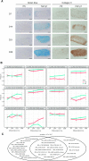
Modulation of human rRNA pseudouridylation profile during TGF-β1 stimulated differentiation of human bone marrow stem cell into chondrocyte-like cells. (A) Accumulation of glycosaminoglycans revealed by alcian blue staining (blue, left) and immunostaining for collagen type II (brown, right). Samples were analyzed at days 7 (D7), 14 (D14), 21 (D21) and 28 (D28). The type of medium used is indicated at the top of each column. For simplicity, only one biological replicate is shown on Figure. (B) Time courses of variations of PsiScore levels at selected pseudouridylated sites in human rRNA. Only highly variable positions are shown, other are presented in Supplementary Figure S11. Differentiation was followed for three independent biological replicates for the BMMSCs issued from the same patient. (C) Human rRNA pseudouridylation sites showing variability between different cell lines and during BMMSCs differentiation. Venn diagram illustrates the overlap of two different datasets.

References

    1. Li X., Xiong X., Yi C.. Epitranscriptome sequencing technologies: decoding RNA modifications. Nat. Methods. 2016; 14:23–31. - PubMed
    1. Helm M., Motorin Y.. Detecting RNA modifications in the epitranscriptome: predict and validate. Nat. Rev. Genet. 2017; 18:275–291. - PubMed
    1. Grozhik A.V., Jaffrey S.R.. Distinguishing RNA modifications from noise in epitranscriptome maps. Nat. Chem. Biol. 2018; 14:215–225. - PubMed
    1. Motorin Y., Helm M.. Methods for RNA modification mapping using deep sequencing: established and new emerging technologies. Genes (Basel). 2019; 10:35. - PMC - PubMed
    1. Harcourt E.M., Kietrys A.M., Kool E.T.. Chemical and structural effects of base modifications in messenger RNA. Nature. 2017; 541:339–346. - PMC - PubMed

Publication types