. 2020 Dec;111(12):4393-4404.
doi: 10.1111/cas.14665. Epub 2020 Oct 7.

NH2 -terminal fragment of ZF21 protein suppresses tumor invasion via inhibiting the interaction of ZF21 with FAK

Affiliations

NH2 -terminal fragment of ZF21 protein suppresses tumor invasion via inhibiting the interaction of ZF21 with FAK

Makoto Nagano et al. Cancer Sci. 2020 Dec.

Abstract

Cellular migration, coupled with the degradation of the extracellular matrix (ECM), is a key step in tumor invasion and represents a promising therapeutic target in malignant tumors. Focal adhesions (FAs) and invadopodia, which are distinct actin-based cellular structures, play key roles in cellular migration and ECM degradation, respectively. The molecular machinery coordinating the dynamics between FAs and invadopodia is not fully understood, although several lines of evidence suggest that the disassembly of FAs is an important step in triggering the formation of invadopodia. In a previous study, we identified the ZF21 protein as a regulator of both FA turnover and invadopodia-dependent ECM degradation. ZF21 interacts with multiple factors for FA turnover, including focal adhesion kinase (FAK), microtubules, m-Calpain, and Src homology region 2-containing protein tyrosine phosphatase 2 (SHP-2). In particular, the dephosphorylation of FAK by ZF21 is a key event in tumor invasion. However, the precise role of ZF21 binding to FAK remains unclear. We established a method to disrupt the interaction between ZF21 and FAK using the FAK-binding NH2 -terminal region of ZF21. Tumor cells expressing the ZF21-derived polypeptide had significantly decreased FA turnover, migration, invadopodia-dependent ECM degradation, and Matrigel invasion. Furthermore, the expression of the polypeptide inhibited an early step of experimental lung metastasis in mice. These findings indicate that the interaction of ZF21 with FAK is necessary for FA turnover as well as ECM degradation at the invadopodia. Thus, ZF21 is a potential regulator that coordinates the equilibrium between FA turnover and invadopodia activity by interacting with FAK.

Keywords: ZF21 protein; extracellular matrix; focal adhesion kinase; invadopodia; tumor metastasis.


Conflict of interest statement

The authors have no conflict of interest.

Figures

FIGURE 1
FIGURE 1
Inhibitory effect of ZF21NT fragment on ZF21 binding to FAK. A, Model proposed for ZF21NT‐mediated tumor suppression, in which ZF21NT inhibits the interaction between FAK and ZF21. B, Purification of ZF21 or EGFP derivatives used for the pull‐down assays. (Left) Schematic representation of ZF21 or EGFP derivatives used for the pull‐down assays in (C). GST, glutathione‐S‐transferase tag; 6× His, hexa‐histidine tag; ZF21NT, N‐terminal 1‐105 aa residues of ZF21 protein. (Right) Coomassie Brilliant Blue (CBB) staining of the purified recombinant proteins. Arrows indicate intact molecules of ZF21 or EGFP derivatives. Asterisk indicates the degraded form. 1, GST‐EGFP; 2, GST‐ZF21; 3, 6× His‐EGFP; 4, 6× His‐ZF21NT. C, Effect of ZF21NT on the interaction of FAK with ZF21
FIGURE 2
FIGURE 2
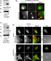
Effect of ZF21NT fragment on the ZF21 localization at FAs. A, Expression levels of m1Venus‐tagged ZF21NT fragments in the cells. B, Colocalization of m1V‐ZF21NT with mCherry‐tagged FAK in the cells. Boxed areas are shown at a higher magnification below. Yellow arrowheads indicate the colocalization of ZF21NT and FAK. C, Expression levels of the V5‐tagged ZF21NT fragment in the ZF21‐m1Venus (ZF21‐m1V)‐expressing cells. D, After 48 h, the localization of ZF21‐m1V and mCherry‐FAK at the cell‐ECM adhesion sites was specifically visualized by total internal reflection fluorescence microscopy (TIRFM). Intracellular distribution of ZF21‐m1V was visualized under the epifluorescence microscope (Epi). Boxed areas are shown at a higher magnification below. Yellow arrowheads indicate the colocalization of ZF21 and FAK. Scale bar, 10 μm
FIGURE 3
FIGURE 3
Effect of ZF21NT fragment on the microtubule‐dependent FAK dephosphorylation. A, Dephosphorylation assay of FAK phosphorylated at Tyr397. B, Phosphatase dependency of FAK dephosphorylation at Tyr397 in ZF21NT expressing cells. C, SHP‐2 dependency of FAK dephosphorylation at Tyr397 in ZF21NT expressing cells
FIGURE 4
FIGURE 4
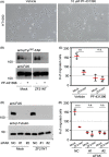
Effect of ZF21NT fragment on the migratory activity of the cells. A, Effect of the FAK inhibitor, PF‐431396, on the viability of HT1080 cells. B, Phosphorylation levels of FAK at Tyr397 in the presence of PF‐431396. C, Effect of FAK inhibitor on the migratory activity of ZF21NT expressing cells. D, Expression levels of FAK in siFAK‐treated cells. E, Effect of FAK knockdown on the migratory activity of ZF21NT expressing cells. Data show mean ± SEM from 3 independent experiments. ***P < .005, ****P < .0001, 2‐way ANOVA with Tukey post hoc test. Scale bars, 250 μm
FIGURE 5
FIGURE 5
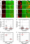
Effect of ZF21NT fragment on the ECM‐degrading activity at the invadopodia. A, C, ECM‐degrading activity of V5 epitope‐tagged ZF21NT expressing cells, HT1080 (A), or MDA‐MB231 (C), stained with phalloidin for localization of filamentous actin (actin panels). Degraded ECM appears as dark areas (OG‐gelatin panels). The boxed areas are shown at higher magnification in the lower panels (boxed area). Yellow or magenta arrowheads indicate the actin‐rich structures or ECM‐degraded spots, respectively. B, D, Quantification of ECM degradation area at the invadopodia structures. Average degradation area per cell in (A) or (C) was calculated and presented in (B) or (D), respectively. E, F, Invasive activity of ZF21NT expressing cells, HT1080 (E) or MDA‐MB231 (F). Data show mean ± SD with at least 100 actin‐rich spots (B, D) or mean ± SEM from 3 independent experiments (E, F). **P < .01, ****P < .0001, unpaired t test with Welch correction. Scale bar, 10 μm
FIGURE 6
FIGURE 6
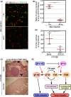
Effect of ZF21NT fragment on lung metastasis in mice model. A, Lung metastatic colonization assay in mouse xenograft model. B, Ratio of lung metastatic colonies formed by ZF21NT cells to mock. The number of green or red fluorescent colonies in (A) was counted. C, H&E staining of murine lungs 28 d after injection of control or ZF21NT cells into the tail veins of 8‐wk‐old nude mice. D, The numbers of tumor nodules in (C). E, Role of ZF21 and effect of ZF21NT in tumor invasion. Data show mean ± SEM with 6 (B) or 5 (D) lung tissues. **P < .01, ****P < .0001, unpaired t test with Welch correction. Scale bar, 200 μm
FIGURE 7
FIGURE 7
Minimizing the ZF21‐derived polypeptide having an inhibitory effect on tumor invasion. A, C, Region of ZF21NT‐fragmented polypeptide using a bait for the FAK‐binding assay. B, D, Pull‐down assay of FAK using GST‐ZF21 derivatives. (Upper) FAK bound to GST‐ZF21 was analyzed by western blotting using the anti‐FAK antibody. (Lower) CBB staining of the bait proteins. E, Expression levels of the m1Venus‐tagged ZF21(41‐75) fragment in HT1080 cells. F, Invasive activity of ZF21(41‐75)‐expressing cells. G, Cell proliferation activity of ZF21(41‐75)‐expressing cells. The data show the mean ± standard error of the mean from 3 independent experiments (F, G). *P < .05, unpaired t test with Welch correction

References

    1. Friedl P, Wolf K. Tumour‐cell invasion and migration: diversity and escape mechanisms. Nat Rev Cancer. 2003;3:362‐374. - PubMed
    1. Gupta GP, Massague J. Cancer metastasis: building a framework. Cell. 2006;127:679‐695. - PubMed
    1. Steeg PS. Tumor metastasis: mechanistic insights and clinical challenges. Nat Med. 2006;12:895‐904. - PubMed
    1. Wehrle‐Haller B, Imhof B. The inner lives of focal adhesions. Trends Cell Biol. 2002;12:382‐389. - PubMed
    1. Zamir E, Geiger B. Molecular complexity and dynamics of cell‐matrix adhesions. J Cell Sci. 2001;114:3583‐3590. - PubMed

Substances