. 2020 Oct 26;60(10):4894-4903.
doi: 10.1021/acs.jcim.0c00589. Epub 2020 Oct 6.

PRosettaC: Rosetta Based Modeling of PROTAC Mediated Ternary Complexes

Affiliations

PRosettaC: Rosetta Based Modeling of PROTAC Mediated Ternary Complexes

Daniel Zaidman et al. J Chem Inf Model. .

Abstract

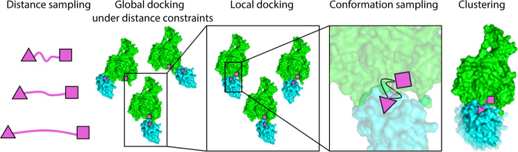
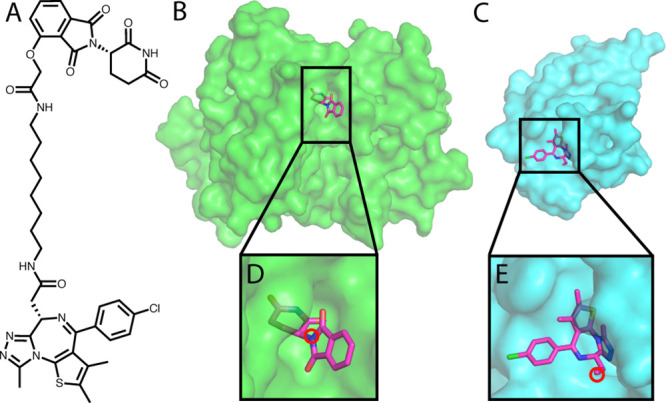
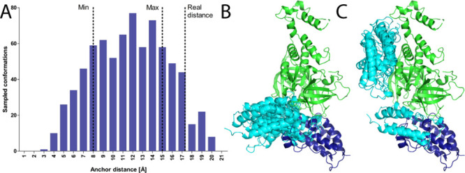
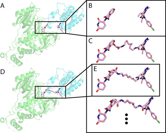
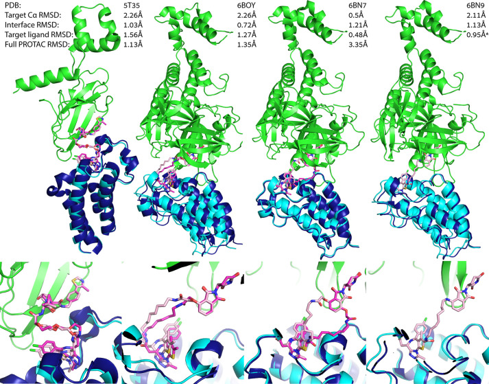
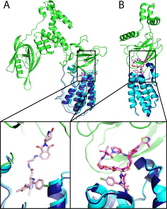
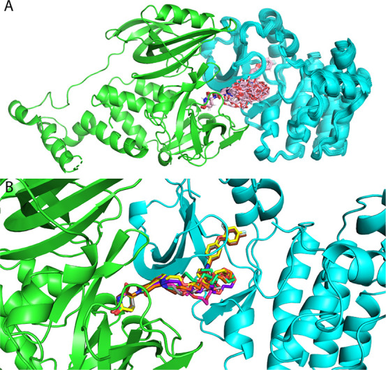
Proteolysis-targeting chimeras (PROTACs), which induce degradation by recruitment of an E3 ligase to a target protein, are gaining much interest as a new pharmacological modality. However, designing PROTACs is challenging. Formation of a ternary complex between the protein target, the PROTAC, and the recruited E3 ligase is considered paramount for successful degradation. A structural model of this ternary complex could in principle inform rational PROTAC design. Unfortunately, only a handful of structures are available for such complexes, necessitating tools for their modeling. We developed a combined protocol for the modeling of a ternary complex induced by a given PROTAC. Our protocol alternates between sampling of the protein-protein interaction space and the PROTAC molecule conformational space. Application of this protocol-PRosettaC-to a benchmark of known PROTAC ternary complexes results in near-native predictions, with often atomic accuracy prediction of the protein chains, as well as the PROTAC binding moieties. It allowed the modeling of a CRBN/BTK complex that recapitulated experimental results for a series of PROTACs. PRosettaC generated models may be used to design PROTACs for new targets, as well as improve PROTACs for existing targets, potentially cutting down time and synthesis efforts. To enable wide access to this protocol, we have made it available through a web server (https://prosettac.weizmann.ac.il/).


Conflict of interest statement

The authors declare no competing financial interest.

Figures

Figure 1
Figure 1
An overview of the PRosettaC protocol. The protocol consists of the following consecutive steps: 1. Sampling of the distance between the two ligand anchor points. 2. Constrained global protein–protein docking with PatchDock. 3. Local docking with RosettaDock. 4. Generating constrained PROTAC conformations compatible with the local docking solutions. 5. Clustering of the top scoring results.
Figure 2
Figure 2
The input for the PRosettaC protocol. A. The chemical structure of a BRD4 PROTAC molecule including a CRBN ligand (thalidomide), a BRD4 ligand (JQ1), and an alkane linker. B. Structure of CRBN with thalidomide. C. Structure of BRD4 with JQ1. D. Thalidomide with the anchor atom marked. E. JQ1 with the anchor atom marked. All structures are based on PDB: 6BOY.
Figure 3
Figure 3
Sampling the PROTAC anchor points distance distribution successfully constrains global docking. A. For each distance (in 1 Å increments), we generate 200 random orientations of the target and E3 ligands and try to generate PROTAC conformations to match these random orientations. The histogram represents successfully generated conformations of an example PROTAC (PDB: 6BOY). On the basis of this histogram, the constraints chosen for the following step were 8—15 Å. Despite the crystallographic distance falling outside the constraints boundaries in this example, the protocol eventually is able to recapitulate the correct binding mode. See Figure S1 and Table S1 for more details on boundary selection and the crystallographic distances. B. Top 5 solutions of global protein–protein docking, constrained by the range calculated by the previous step of the protocol. C. Top 5 solutions of unconstrained protein–protein docking. Green, E3 ligase CRBN; dark blue, crystallographic BRD4 (6BOY); cyan, docked BRD4.
Figure 4
Figure 4
Generating PROTAC conformations in the context of the protein–protein interaction. Using RDKit, we generate up to 100 PROTAC conformations, to match the ligand orientation of each local docking solution. Then, we use Rosetta Packer to choose the best conformation to fit the protein–protein docking model. A. An example local docking solution for 6BOY. B. The ligand orientation, extracted from the local docking solution. C. Constrained conformations that bridge between the ligand orientation. D. Output model of Rosetta after choosing the best constrained conformation. E. The conformation chosen by Rosetta. Green, E3 ligase CRBN; cyan, predicted BRD4; light pink, predicted PROTAC conformation.
Figure 5
Figure 5
PRosettaC can recapitulate known ternary complexes to atomic accuracy. Upper panels: near native predictions for four structures of the training set: 5T35, 6BOY, 6BN7 and 6BN9, with reported Cα RMSD of the moving chain (BRD4), protein interface RMSD which is calculated on all backbone atoms of residues which are within 8 Å of any residue on the other protein, the RMSD over the small molecule ligands (excluding the linker), and the RMSD over the entire PROTAC. Lower panels: zoom in on the ligand binding predictions. In all four, PRosettaC achieved predictions of atomic accuracy for the target ligands. Green, E3 ligase CRBN; dark blue, X-ray BRD4; cyan, docked BRD4; magenta, X-ray PROTAC conformation; light pink, predicted PROTAC conformation. *Note that the PROTAC molecule is not resolved in 6BN9 and was modeled by PRosettaC. The RMSD values for the ligands are based on alignment to 6BN7. Since the ligand–protein interactions are highly conserved, the ligand RMSD, based on the alignment, should still be representative.
Figure 6
Figure 6
Near native predictions for two nontraditional PROTAC ternary complexes. A. Near native prediction of PDB: 6BNB of a BRD4 PROTAC with an open conformation of CRBN. The target Cα RMSD is 2.35 Å. The native cluster was ranked 1, containing 67.7% of the final models. The PROTAC molecule is not modeled in the original structure, but could be easily modeled in using ProsettaC. B. Near native prediction of 6SIS, a complex of BRD4 and CRBN with a macrocyclic PROTAC. The target Cα RMSD is 1.3 Å, with a target ligand RMSD of 0.5 Å and a full PROTAC RMSD of 0.72 Å. The native cluster was ranked 3. Green, E3 ligase; dark blue, crystallographic BRD4; cyan, predicted BRD4; magenta, X-ray PROTAC conformation; light pink, predicted PROTAC conformation.
Figure 7
Figure 7
High confidence prediction of a BTK–CRBN ternary complex. Modeling of a BTK–CRBN ternary complex, with a series of 11 PROTACs, recapitulates which PROTACs are active and which are not and suggests a high-confidence model for the interaction. A. A cluster (with 0.5 Å threshold) of 33 models from various PRosettaC runs contains representatives from active PROTACs 5–11. B. Representative of PROTACs 5–11: 5, brown; 6, gray; 7, magenta; 8, yellow; 9, orange; 10, purple; 11, green.

References

    1. Toure M.; Crews C. M. Small-Molecule PROTACS: New Approaches to Protein Degradation. Angew. Chem., Int. Ed. 2016, 55, 1966–1973. 10.1002/anie.201507978. - DOI - PubMed
    1. Neklesa T. K.; Winkler J. D.; Crews C. M. Targeted Protein Degradation by PROTACs. Pharmacol. Ther. 2017, 174, 138–144. 10.1016/j.pharmthera.2017.02.027. - DOI - PubMed
    1. Pettersson M.; Crews C. M. Proteolysis TArgeting Chimeras (PROTACs)—past, Present and Future. Drug Discovery Today: Technol. 2019, 31, 15.10.1016/j.ddtec.2019.01.002. - DOI - PMC - PubMed
    1. Gadd M. S.; Testa A.; Lucas X.; Chan K.-H.; Chen W.; Lamont D. J.; Zengerle M.; Ciulli A. Structural Basis of PROTAC Cooperative Recognition for Selective Protein Degradation. Nat. Chem. Biol. 2017, 13, 514–521. 10.1038/nchembio.2329. - DOI - PMC - PubMed
    1. Smith B. E.; Wang S. L.; Jaime-Figueroa S.; Harbin A.; Wang J.; Hamman B. D.; Crews C. M. Differential PROTAC Substrate Specificity Dictated by Orientation of Recruited E3 Ligase. Nat. Commun. 2019, 10, 131.10.1038/s41467-018-08027-7. - DOI - PMC - PubMed

Publication types

MeSH terms

Substances