An official website of the United States government
The .gov means it’s official.
Federal government websites often end in .gov or .mil. Before
sharing sensitive information, make sure you’re on a federal
government site.
The site is secure.
The https:// ensures that you are connecting to the
official website and that any information you provide is encrypted
and transmitted securely.
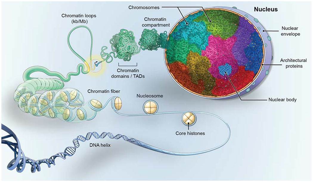
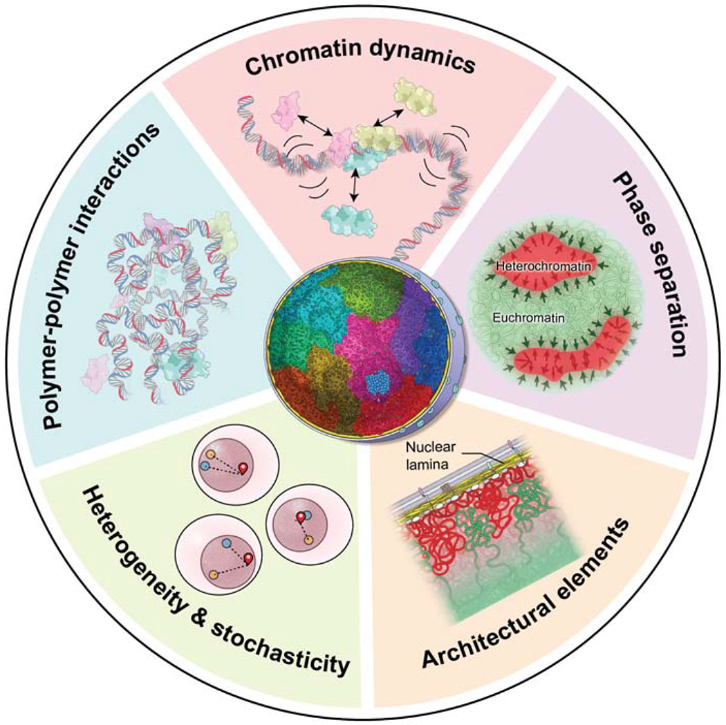
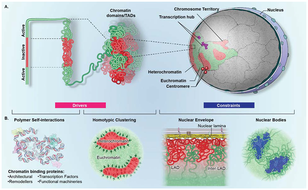
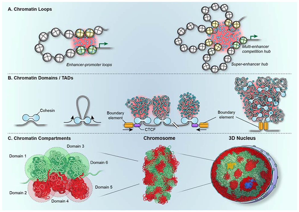
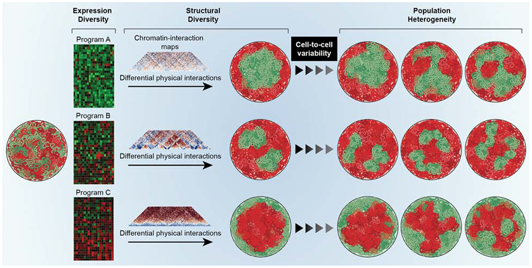
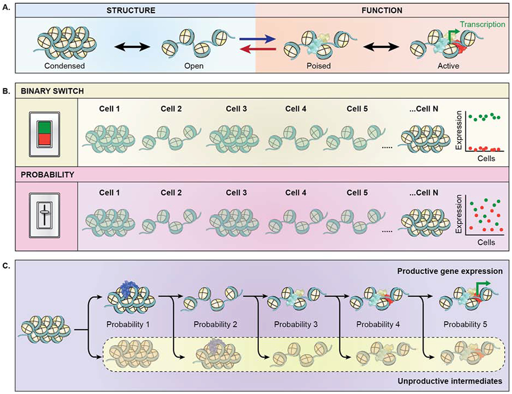
Genomes have complex three-dimensional architectures. The recent convergence of genetic, biochemical, biophysical, and cell biological methods has uncovered several fundamental principles of genome organization. They highlight that genome function is a major driver of genome architecture and that structural features of chromatin act as modulators, rather than binary determinants, of genome activity. The interplay of these principles in the context of self-organization can account for the emergence of structural chromatin features, the diversity and single-cell heterogeneity of nuclear architecture in cell types and tissues, and explains evolutionarily conserved functional features of genomes, including plasticity and robustness.
Figure 1.. The organization of the eukaryotic genome.
Genomes are organized at multiple levels. DNA…
Figure 1.. The organization of the eukaryotic genome.
Genomes are organized at multiple levels. DNA is wrapped around the nucleosome, which is made up of an octamer of core-histones, forming the chromatin fiber which folds into loops, often bringing upstream gene regulatory elements (yellow), such as enhancers, into proximity to genes (blue) to control their transcription (black arrow). The fiber then folds into chromatin domains, referred to as topologically associating domains (TADs), which associate with each other to create chromatin compartments. The DNA of each chromosome occupies a distinct volume, or chromosome territory (multiple colors), within the cell nucleus, generating non-random patterns of chromosome and gene locations. In the DNA-free space, the nucleus also contains RNA and proteinaceous protein aggregates which form nuclear bodies (blue).
Figure 2.. Principles of genome organization.
The…
Figure 2.. Principles of genome organization.
The interplay of several fundamental principles governs genome organization.…
Figure 2.. Principles of genome organization.
The interplay of several fundamental principles governs genome organization. (Light blue sector) Polymer-polymer interactions, mediated by chromatin binding proteins (light blue, pink, yellow), promote the formation of chromatin loops and shape the overall conformation of the chromatin fiber. (Pink sector) Chromatin undergoes local motion and chromatin-chromatin interactions are transient. Chromatin proteins (light blue, pink, yellow) undergo rapid cycles of association and dissociation with short residence times. (Purple sector) The process of phase separation involves the homotypic aggregation of proteins (arrows) and contributes to the formation and stabilization of chromatin-chromatin interactions and domains, including eu- and heterochromatin. (Orange sector) The physical interaction of chromatin with stable architectural elements of the nucleus such as the nuclear envelope limits the degree of freedom of a genome region and contributes to its non-random localization. (Green sector) the behavior of individual chromatin regions and genes (red, blue, yellow) varies stochastically in individual cells giving rise to extensive heterogeneity in genome organization and function amongst single cells in a population.
Figure 3.. Drivers and constraints in genome…
Figure 3.. Drivers and constraints in genome organization.
(A) Genome organization is determined by the…
Figure 3.. Drivers and constraints in genome organization.
(A) Genome organization is determined by the interplay of drivers (pink box) and constraints (blue box). Higher-order organization is generated by self-interaction of transcriptionally active (green) and inactive (red) genome regions which fold onto themselves to form domains. Multiple homotypic (active or inactive) domains in turn aggregate both within a single chromosome and between domains on distinct chromosomes to form compartments. Homotypic interactions amongst multiple chromosomes generate transcription hubs (purple) and centromere aggregates (red spheres) which contain genes and centromeres located on distinct chromosomes, respectively. (B) (left) Drivers of genome organization include polymer-polymer interactions and phase separation. Homotypic polymer-polymer interactions among chromatin fibers form domains that are stabilized by phase separation (black arrows), including segregation of eu- and heterochromatin. (right) Constraints of genome organization include architectural features that physically interact with genome regions to influence their mobility and location, such as the nuclear lamina, which interacts with transcriptionally inactive lamina-associated domains (LADs) (red), interrupted by inter-LADs (green) or nuclear bodies, such as splicing speckles (blue), which preferentially associate with transcriptionally active chromatin.
Figure 4.. Formation of chromatin features by…
Figure 4.. Formation of chromatin features by self-organization.
(A) Chromatin loops, including promoter-enhancer loops form…
Figure 4.. Formation of chromatin features by self-organization.
(A) Chromatin loops, including promoter-enhancer loops form through the close physical proximity of genome regions. Their interactions are stabilized by the formation of phase-separated aggregates (shaded in red) of relevant chromatin proteins and transcription factors (blue squares). Multiple enhancers (yellow nucleosomes) may associate with the same gene promoter (green nucleosomes) to activate the same gene target (green arrow) in multi-enhancer hubs or in superenhancers. (B) TADs form via loop-extrusion generated by the cohesin motor (light blue). The CTCF protein (purple) defines the boundary of the domain (gold) by determining the location of cohesin. The internal structure of TADs is determined by heterogenous polymer chromatin-chromatin interactions, which are likely stabilized by phase separation (shaded in red). Black arrows denote the direction of movement of DNA through the cohesin complex. (C) (left, middle) Chromatin compartments form via the association of multiple homotypic domains (1-6; green and red) to ultimately form chromosomes. (middle, right) Multiple chromosomes associate via interactions between homotypic domains across the 3D structure of the nucleus to form large-scale blocks of heterochromatin (red) and euchromatin (green). Nuclear bodies (blue) serve as anchoring points. Transcription hubs (yellow) form due to the coalescence of multiple active genome regions located on multiple chromosomes.
Figure 5.. Genome function drives structure.
All…
Figure 5.. Genome function drives structure.
All cells of an organism contain genomes of identical…
Figure 5.. Genome function drives structure.
All cells of an organism contain genomes of identical sequence (left single nucleus), but different cell-types express distinct gene expression programs (heatmaps) and consequently have different genome topologies as detected by chromatin interaction maps. The cell-type specific homotypic chromatin-chromatin interactions drive higher-order genome organization and generate distinct overall genome topologies in different cell types, resulting in cell-type specific patterns of euchromatin (green) and heterochromatin (red). (right) Within a cell type, the heterogeneity of chromatin-chromatin interactions generates cell-to-cell variability in the population.
Figure 6.. The probabilistic genome.
(A) Bi-directional…
Figure 6.. The probabilistic genome.
(A) Bi-directional interplay of chromatin structure (blue shaded area) and…
Figure 6.. The probabilistic genome.
(A) Bi-directional interplay of chromatin structure (blue shaded area) and function (orange shaded area). Chromatin structure affects gene function (blue forward arrow). Chromatin structure reversibly oscillates between a condensed and open state, which facilitates association of transcription factors (blue, yellow), leading to a poised stated and upon association of RNA polymerase (red) enables transcription (green bent arrow). Conversely, transcription affects structure (red reverse arrow), by maintaining an open chromatin structure. (B) Chromatin structure does not act as a deterministic, binary switch, but rather as a probabilistic modulator of function. (green panel) In a binary switch model, the expression level of a given gene in individual cells is either fully on (green points) or fully off (red points) reflecting the open and closed configuration of the gene locus in single cells. (pink panel) In a modulatory model, gene expression levels are heterogeneous (red, green points) in individual cells reflecting the probabilistic nature of gene activity in open and closed chromatin. Variable expression levels are typically observed experimentally. (C) Gene expression is a multi-step processes and is inherently probabilistic. Each step required to activate a gene represents an equilibrium of a productive event towards gene activation versus an unproductive event with a certain probability. Probability 1: stable association of a chromatin remodeling factors (blue) vs. transient interaction as they diffuse through the nucleus. Probability 2: maintenance of decondensed chromatin vs. reversion to condensed state. Probability 3: association of early transcription factors (light blue, yellow) which promotes the likelihood of association of additional transcription factors vs. disassociation of early transcription factor. Probability 4: association of RNA polymerase (red) vs. loss of activating transcription factors. Probability 5: transcriptional activation (bent green arrow) vs. unproductive dissociation of RNA polymerase. As a result of the probabilistic nature of each step, the activation process as a whole is relatively inefficient, thus generating the stochastic patterns of activation observed for most genes.
Banani SF, Lee HO, Hyman AA, and Rosen MK (2017). Biomolecular condensates: organizers of cellular biochemistry. Nat Rev Mol Cell Biol 18, 285–298.
-
PMC
-
PubMed
Bar-Even A, Paulsson J, Maheshri N, Carmi M, O’Shea E, Pilpel Y, and Barkai N (2006). Noise in protein expression scales with natural protein abundance. Nat Genet 38, 636–643.
-
PubMed
Barbieri M, Chotalia M, Fraser J, Lavitas LM, Dostie J, Pombo A, and Nicodemi M (2012). Complexity of chromatin folding is captured by the strings and binders switch model. Proc Natl Acad Sci U S A 109, 16173–16178.
-
PMC
-
PubMed
Beagrie RA, Scialdone A, Schueler M, Kraemer DC, Chotalia M, Xie SQ, Barbieri M, de Santiago I, Lavitas LM, Branco MR, et al. (2017). Complex multi-enhancer contacts captured by genome architecture mapping. Nature 543, 519–524.
-
PMC
-
PubMed
Bertero A, Fields PA, Ramani V, Bonora G, Yardimci GG, Reinecke H, Pabon L, Noble WS, Shendure J, and Murry CE (2019). Dynamics of genome reorganization during human cardiogenesis reveal an RBM20-dependent splicing factory. Nat Commun 10, 1538.
-
PMC
-
PubMed