

Structural white and gray matter differences in a large sample of patients with Posttraumatic Stress Disorder and a healthy and trauma-exposed control group: Diffusion tensor imaging and region-based morphometry
- PMID: 32977211
- PMCID: PMC7511745
- DOI: 10.1016/j.nicl.2020.102424
Structural white and gray matter differences in a large sample of patients with Posttraumatic Stress Disorder and a healthy and trauma-exposed control group: Diffusion tensor imaging and region-based morphometry
Abstract
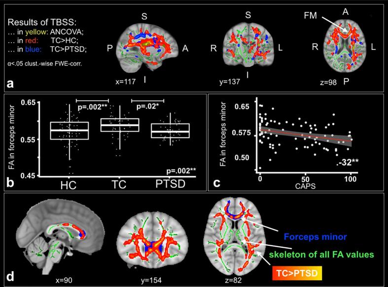
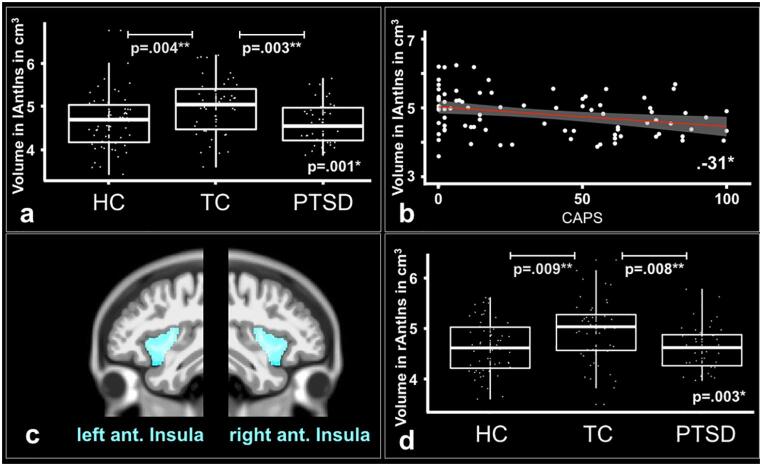
Differences in structural white and gray matter in survivors of traumatic experiences have been related to the development and maintenance of Posttraumatic Stress Disorder (PTSD). However, there are very few studies on diffusion tensor imaging and region based morphometry comparing patients with PTSD to two control groups, namely healthy individuals with or without trauma experience. It is also unknown if differences in white and gray matter are associated. In this cross-sectional study, we examined white- and gray matter differences between 44 patients with PTSD, 49 trauma control and 61 healthy control subjects. We compared the groups applying Tract-Based Spatial Statistics (TBSS) for a whole brain white matter analysis as well as region of interest analyses for white and gray matter. First, trauma control subjects in comparison to patients with PTSD and healthy control subjects showed significantly a) higher fractional anisotropy (FA) in the left corticospinal tract and inferior fronto-occipital fasciculus than patients with PTSD, b) higher FA in the left inferior fronto-occipital-, right inferior- and right superior longitudinal fasciculi, c) higher FA in the forceps minor and d) higher volume of the left and right anterior insulae. Second, we show significant correlations between the FA in the forceps minor and the gray matter volume in the left and right anterior insulae. Third, the mean FA value in the forceps minor correlated negatively with symptom severity of PTSD and depression as well as trait anxiety, whereas the gray matter volume in the left anterior insula correlated negatively with symptom severity in PTSD. Our findings underline the importance of brain structures critically involved in emotion regulation and salience mapping. While previous studies associated these processes primarily to functional and task-based differences in brain activity, we argue that morphometrical white and gray matter differences could serve as targets in neuroscientifically-informed prevention and treatment interventions for PTSD.
Keywords: Diffusion tensor imaging; Neuroplasticity; PTSD; Trauma; White and gray matter.
Copyright © 2020 The Author(s). Published by Elsevier Inc. All rights reserved.
Conflict of interest statement
The authors declare that they have no known competing financial interests or personal relationships that could have appeared to influence the work reported in this paper.
Figures

Similar articles
-
Association of abnormal white matter integrity in the acute phase of motor vehicle accidents with post-traumatic stress disorder.J Affect Disord. 2016 Jan 15;190:714-722. doi: 10.1016/j.jad.2015.09.044. Epub 2015 Nov 5. J Affect Disord. 2016. PMID: 26600413
-
Disruption of white matter structural integrity and connectivity in posttraumatic stress disorder: A TBSS and tractography study.Depress Anxiety. 2017 May;34(5):437-445. doi: 10.1002/da.22615. Epub 2017 Mar 15. Depress Anxiety. 2017. PMID: 28294462 Free PMC article.
-
[A study on longitudinal changes in white matter microstructure of parents who have lost their only child based on diffusion tensor imaging and its relationship with symptoms of posttraumatic stress disorder].Zhonghua Yi Xue Za Zhi. 2022 Jun 21;102(23):1760-1765. doi: 10.3760/cma.j.cn112137-20211213-02778. Zhonghua Yi Xue Za Zhi. 2022. PMID: 35705480 Chinese.
-
White matter microstructural alterations in posttraumatic stress disorder: An ROI and whole-brain based meta-analysis.J Affect Disord. 2020 Apr 1;266:655-670. doi: 10.1016/j.jad.2020.01.047. Epub 2020 Jan 16. J Affect Disord. 2020. PMID: 32056942 Review.
-
White matter microstructural abnormalities and gray matter volume alterations in obsessive-compulsive disorder: A coordinate-based meta-analysis.J Affect Disord. 2023 Jan 1;320:751-761. doi: 10.1016/j.jad.2022.09.035. Epub 2022 Sep 26. J Affect Disord. 2023. PMID: 36174788 Review.
Cited by
-
Neurostructural associations with traumatic experiences during child- and adulthood.Transl Psychiatry. 2022 Dec 15;12(1):515. doi: 10.1038/s41398-022-02262-9. Transl Psychiatry. 2022. PMID: 36517466 Free PMC article.
-
Low levels of Methyl-CpG binding protein 2 are accompanied by an increased vulnerability to the negative outcomes of stress exposure during childhood in healthy women.Transl Psychiatry. 2022 Dec 8;12(1):506. doi: 10.1038/s41398-022-02259-4. Transl Psychiatry. 2022. PMID: 36481643 Free PMC article.
-
Psychoradiological abnormalities in treatment-naive noncomorbid patients with posttraumatic stress disorder.Depress Anxiety. 2022 Jan;39(1):83-91. doi: 10.1002/da.23226. Epub 2021 Nov 18. Depress Anxiety. 2022. PMID: 34793618 Free PMC article.
-
Cross-sectional field study comparing hippocampal subfields in patients with post-traumatic stress disorder, major depressive disorder, post-traumatic stress disorder with comorbid major depressive disorder, and adjustment disorder using routine clinical data.Front Psychol. 2023 Jun 13;14:1123079. doi: 10.3389/fpsyg.2023.1123079. eCollection 2023. Front Psychol. 2023. PMID: 37384185 Free PMC article.
-
Assessing biological self-organization patterns using statistical complexity characteristics: a tool for diffusion tensor imaging analysis.MAGMA. 2024 Jul 28. doi: 10.1007/s10334-024-01185-4. Online ahead of print. MAGMA. 2024. PMID: 39068635
References
-
- American Psychiatric Association. (2000). Diagnostic and Statistical Manual of Mental Disorders, Fourth Edition, Text Revision (DSM-IV-TR) (Vol. 1). https://doi.org/10.1176/appi.books.9780890423349.
-
- American Psychiatric Association. (2013). Guía de consulta de los criterios diagnósticos del DSM-5®. https://doi.org/10.1176/appi.books.9780890425657.
Publication types
MeSH terms
LinkOut - more resources
Full Text Sources
Medical
