

Neurology of COVID-19 in Singapore
- PMID: 32977228
- PMCID: PMC7470792
- DOI: 10.1016/j.jns.2020.117118
Neurology of COVID-19 in Singapore
Erratum in
-
Corrigendum to 'Neurology of COVID-19 in Singapore' [Journal of the Neurological Sciences Volume 418, 15 November 2020, 117118].J Neurol Sci. 2021 May 15;424:117406. doi: 10.1016/j.jns.2021.117406. Epub 2021 Mar 20. J Neurol Sci. 2021. PMID: 33773769 Free PMC article. No abstract available.
Abstract
Purpose: To describe the spectrum of COVID-19 neurology in Singapore.
Method: We prospectively studied all microbiologically-confirmed COVID-19 patients in Singapore, who were referred for any neurological complaint within three months of COVID-19 onset. Neurological diagnoses and relationship to COVID-19 was made by consensus guided by contemporaneous literature, refined using recent case definitions.
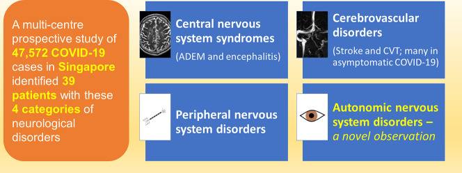
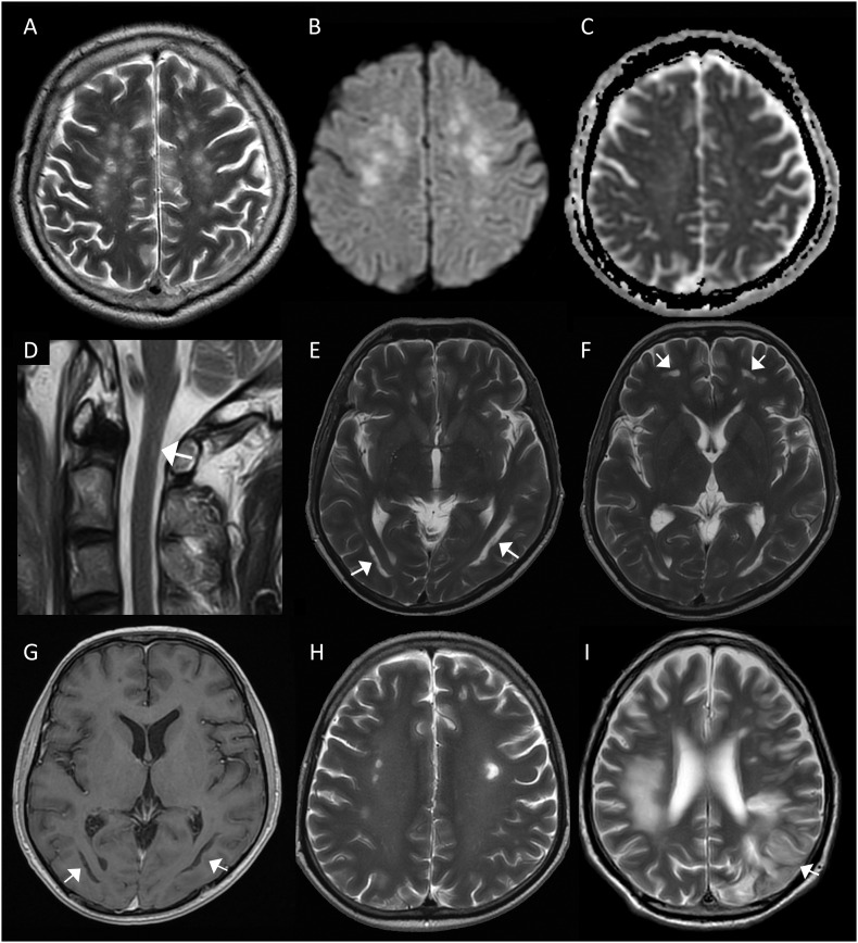
Results: 47,572 patients (median age 34 years, 98% males) were diagnosed with COVID-19 in Singapore between 19 March to 19 July 2020. We identified 90 patients (median age 38, 98.9% males) with neurological disorders; 39 with varying certainty of relationship to COVID-19 categorised as: i) Central nervous system syndromes-4 acute disseminated encephalomyelitis (ADEM) and encephalitis, ii) Cerebrovascular disorders-19 acute ischaemic stroke and transient ischaemic attack (AIS/TIA), 4 cerebral venous thrombosis (CVT), 2 intracerebral haemorrhage, iii) Peripheral nervous system-7 mono/polyneuropathies, and a novel group, iv) Autonomic nervous system-4 limited dysautonomic syndromes. Fifty-one other patients had pre/co-existent neurological conditions unrelated to COVID-19. Encephalitis/ADEM is delayed, occurring in critical COVID-19, while CVT and dysautonomia occurred relatively early, and largely in mild infections. AIS/TIA was variable in onset, occurring in patients with differing COVID-19 severity; remarkably 63.2% were asymptomatic. CVT was more frequent than expected and occurred in mild/asymptomatic patients. There were no neurological complications in all 81 paediatric COVID-19 cases.
Conclusion: COVID-19 neurology has a wide spectrum of dysimmune-thrombotic disorders. We encountered relatively few neurological complications, probably because our outbreak involved largely young men with mild/asymptomatic COVID-19. It is also widely perceived that the pandemic did not unduly affect the Singapore healthcare system.
Keywords: COVID-19; Coronavirus; Neurological manifestations; Pandemic; SARS-CoV-2.
Copyright © 2020 Elsevier B.V. All rights reserved.
Conflict of interest statement
None.
Figures
References
-
- Prerna A., Lim J.Y.X., Tan N.W.H. Neurology of the H1N1 pandemic in Singapore: a nationwide case series of children and adults. J. Neuro-Oncol. 2015;21(5):491–499. - PubMed
-
- World Health Organization . WHO; 2020. The Coronavirus Disease 2019 (COVID-19):Situation Report-36.
MeSH terms
LinkOut - more resources
Full Text Sources
Medical
Miscellaneous
