

Differentiation of Endometriomas from Ovarian Hemorrhagic Cysts at Magnetic Resonance: The Role of Texture Analysis
- PMID: 32977428
- PMCID: PMC7598287
- DOI: 10.3390/medicina56100487
Differentiation of Endometriomas from Ovarian Hemorrhagic Cysts at Magnetic Resonance: The Role of Texture Analysis
Abstract
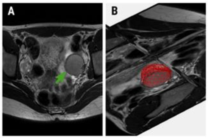
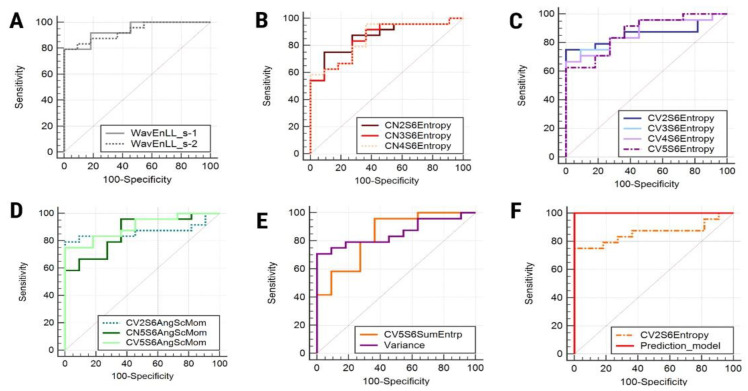
Background and Objectives: To assess ovarian cysts with texture analysis (TA) in magnetic resonance (MRI) images for establishing a differentiation criterion for endometriomas and functional hemorrhagic cysts (HCs) that could potentially outperform their classic MRI diagnostic features. Materials and Methods: Forty-three patients with known ovarian cysts who underwent MRI were retrospectively included (endometriomas, n = 29; HCs, n = 14). TA was performed using dedicated software based on T2-weighted images, by incorporating the whole lesions in a three-dimensional region of interest. The most discriminative texture features were highlighted by three selection methods (Fisher, probability of classification error and average correlation coefficients, and mutual information). The absolute values of these parameters were compared through univariate, multivariate, and receiver operating characteristic analyses. The ability of the two classic diagnostic signs ("T2 shading" and "T2 dark spots") to diagnose endometriomas was assessed by quantifying their sensitivity (Se) and specificity (Sp), following their conventional assessment on T1-and T2-weighted images by two radiologists. Results: The diagnostic power of the one texture parameter that was an independent predictor of endometriomas (entropy, 75% Se and 100% Sp) and of the predictive model composed of all parameters that showed statistically significant results at the univariate analysis (100% Se, 100% Sp) outperformed the ones shown by the classic MRI endometrioma features ("T2 shading", 75.86% Se and 35.71% Sp; "T2 dark spots", 55.17% Se and 64.29% Sp). Conclusion: Whole-lesion MRI TA has the potential to offer a superior discrimination criterion between endometriomas and HCs compared to the classic evaluation of the two lesions' MRI signal behaviors.
Keywords: endometrioma; endometriosis; magnetic resonance imaging (MRI); ovarian cysts; texture analysis.
Conflict of interest statement
The authors declare no conflict of interest.
Figures
References
-
- Carnahan M., Fedor J., Agarwal A., Gupta S. Ovarian endometrioma: Guidelines for selection of cases for surgical treatment or expectant management. Expert Rev. Obstet. Gynecol. 2013;8:29–55. doi: 10.1586/eog.12.75. - DOI
MeSH terms
LinkOut - more resources
Full Text Sources
Medical
