. 2020 Sep 23;9(4):50.
doi: 10.3390/antib9040050.

Taking the Hinge off: An Approach to Effector-Less Monoclonal Antibodies

Affiliations

Taking the Hinge off: An Approach to Effector-Less Monoclonal Antibodies

Jamie Valeich et al. Antibodies (Basel). .

Abstract

A variety of Fc domain engineering approaches for abrogating the effector functions of mAbs exists. To address some of the limitations of the current Fc domain silencing approaches, we are exploring a less commonly considered option which relies on the deletion of the hinge. Removal of the hinge domain in humanized IgG1 and IgG4 mAbs obliterates their ability to bind to activating human Fc gamma receptors I and IIIA, while leaving their ability to engage their target antigen intact. Deletion of the hinge also reduces binding to the Fc neonatal receptor, although Fc engineering allows partial recovery of affinity. Engineering of the CH3 domain, stabilizes hinge deleted IgG4s and prevents Fab arm exchange. The faster clearing properties together with the pacified Fc make modality of the hinge deleted mAb an appealing solution for therapeutic and diagnostic applications.

Keywords: Antibody-dependent cellular cytotoxicity (ADCC); Fab arm exchange; Fc neonatal receptor; FcRn; Fcγ receptors; FcγRI; FcγRIII; IgG1; IgG4; effector function; hinge; mAb; monoclonal antibody.


Conflict of interest statement

The authors declare no conflict of interest.

Figures

Figure 1
Figure 1
(a) Three-dimensional representation of the crystal structures of a human IgG1 (PDB 1HZH) and the hinge deleted Mcg Antibody (PDB 1MCO). (b) Sequence alignment of the IgG1 and IgG4 hinge (IgG1 HC, IgG4 HC) and resulting sequence after deletion (IgG1 Δhinge and IgG4 Δhinge).
Figure 2
Figure 2
Biochemical and biophysical characterization of the hinge deleted IgG4 and its corresponding full-length control mAb by (a) SEC-UPLC coupled to UV (A280) and Multi Angle Laser Light Scattering (MALS). The UV elution profiles of the hinge deleted IgG4 molecule (bottom trace) and the corresponding full-length control (top trace) are shown. MALS data show measured molecular masses of 132 (hinge-deleted) and 149 kDa (full-length mAb). (b) Nonreduced CE-SDS coupled to UV (A280). Electropherograms of the buffer control (bottom trace), hinge deleted IgG4 molecule (middle trace) and the corresponding full-length control (top trace) are shown. (c) Differential Scanning Fluorimetry. An overlay of the melting profiles of the hinge deleted IgG4 molecule (orange) and the corresponding full-length control (teal) are shown. Melting temperatures/onset were 54.6/47.9 °C (hinge-deleted) and 62.7/56.6 °C (full-length mAb). (d) Intact reduced LC–MS. Total Ion Counts of the hinge deleted IgG4 molecule heavy chain (bottom trace) and the corresponding heavy chain of the full-length control (top trace) are shown. The peak corresponding to the dominant Fc N-Glycan G0F species is shown.
Figure 3
Figure 3
Biochemical and biophysical characterization of the hinge deleted IgG1 and its corresponding full-length control mAb by (a) SEC-UPLC coupled to UV (A280) and MALS. The UV elution profiles of the hinge deleted IgG1 molecule (bottom trace) and the corresponding full-length control (top trace) are shown. Molecular masses of 140 and 150 kDa were measured by MALS for the hinge deleted molecule and its full-length counterpart. (b) Nonreduced CE-SDS coupled to UV (A280). Electropherograms of the buffer control (bottom trace), hinge deleted IgG1 molecule (middle trace) and the corresponding full-length control (top trace) are shown. (c) Differential Scanning Fluorimetry. An overlay of the melting profiles of the hinge deleted IgG1 molecule (orange) and the corresponding full-length control (teal) are shown. Melting temperatures/onset were 67.8/56.5 °C for the hinge deleted molecule and 71.1/63.2 °C for the full-length control. (d) Intact reduced LC–MS. Total Ion Counts of the hinge deleted IgG1 molecule heavy chain (top trace) and the corresponding heavy chain of the full-length control (bottom trace) are shown. The peak corresponding to the dominant Fc N-Glycan G0F and G1F species are shown.
Figure 4
Figure 4
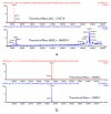
Analysis of the hinge deleted IgG4 and its corresponding full-length control mAb by LC–MS. (a) Deconvoluted spectrum of a nonreduced analysis. Under denaturating conditions, the hinge deleted IgG4 molecule has an apparent mass of 72,161.0 Da consistent with a Heavy-Light (HL) molecule with G0F N-Glycans. The full length mAb control has the expected mass of 146,829.0 Da consistent with the H2L2 structure (with G0F N-Glycans). Under reducing conditions, the heavy chain (Figure 3d) and Light chains (b) can be separated. The hinge deleted IgG4 and the full-length control molecule have the same light chain (23,399.5 Da).
Figure 5
Figure 5
Binding of hinge deleted IgG4 (blue dilution curves) and its corresponding full-length control mAb (green dilution curves) to (a) FcγRI by ELISA, (b) FcRn by ELISA. (c) Enzymatic inhibition of the MMP9 target antigen.
Figure 6
Figure 6
Binding of hinge deleted IgG1 (orange dilution curves) and its corresponding full-length control mAb (blue dilution curves) to (a) FcγRI by competitive AlphaScreen® assay, (b) FcγRIIIa V158 by binding ELISA, (c) FcRn by binding ELISA. Incorporation of point mutations in the Fc of the hinge deleted IgG1 (green dilution series) almost rescues binding to the level of the full-length control. (d) Engagement of the gp120 target antigen by a HEK290 cell-based ELISA reflective of the mAb mode of action.
Figure 7
Figure 7
Blocking Fab arm exchange of hinge deleted mAbs by CH3 stabilization monitored by Förster resonance energy transfer analysis (FAE) which was performed under 1 mM glutathione reducing conditions (a) or in the absence of reducing agent as the negative control (b). Förster resonance energy transfer (FRET) signal (ex 490 nm/Em 515 nm) was recorded for the positive control (Natalizumab x Natalizumab), the hinge deleted IgG4 (Natalizumab x hinge deleted IgG4), the hinge deleted IgG4 with R409K point mutation in the CH3 domain (Natalizumab x R409K hinge deleted IgG4), for the negative control (S228P full length IgG4 x Natalizumab) and for the blank background control. (c) Bar graph summary representing the propensity of each of the test molecules to engage in FAE with Natalizumab.
Figure 8
Figure 8
Blocking Fab arm exchange of hinge deleted mAbs by CH3 stabilization monitored by chromatographic separation. Propensity of IgG4 hinge deleted (T1), R409K CH3 stabilized IgG4 hinge deleted (T2), and S228P hinge stabilized full length IgG4 (WT) to engage in Fab arm exchange with Tysabri® in the presence (a) or the absence (c) of 1 mM GSH reducing agent was analyzed by chromatographic separation. An FAE positive control reaction was conducted by monitoring the formation of the bispecific antibody by chromatographic separation between Tysabri® and a nonhinge stabilized aGFP IgG4 antibody (IgG4 Kappa Antibody) in the presence (b) or the absence (d) of 1 mM GSH reducing agent.

Similar articles

Cited by

References

    1. Vidarsson G., Dekkers G., Rispens T. IgG subclasses and allotypes: From structure to effector functions. Front. Immunol. 2014;5:520. doi: 10.3389/fimmu.2014.00520. - DOI - PMC - PubMed
    1. Bournazos S., Ravetch J.V. Diversification of IgG effector functions. Int. Immunol. 2017;29:303–310. doi: 10.1093/intimm/dxx025. - DOI - PMC - PubMed
    1. Chen Q., Vieth M., Timm D.E., Humblet C., Schneidman-Duhovny D., Chemmama I.E., Sali A., Zeng W., Lu J., Liu L. Reconstruction of 3D structures of MET antibodies from electron microscopy 2D class averages. PLoS ONE. 2017;12:e0175758. doi: 10.1371/journal.pone.0175758. - DOI - PMC - PubMed
    1. Zhang X., Zhang L., Tong H., Peng B., Rames M.J., Zhang S., Ren G. 3D structural fluctuation of IgG1 antibody revealed by individual particle electron tomography. Sci. Rep. 2015;5:9803. doi: 10.1038/srep09803. - DOI - PMC - PubMed
    1. Tian X., Vestergaard B., Thorolfsson M., Yang Z., Rasmussen H.B., Langkilde A.E. In-depth analysis of subclass-specific conformational preferences of IgG antibodies. IUCrJ. 2015;2:9–18. doi: 10.1107/S205225251402209X. - DOI - PMC - PubMed

LinkOut - more resources