

Hepatic epithelioid hemangioendothelioma: case series of a rare vascular tumor mimicking metastases
- PMID: 32977811
- PMCID: PMC7519523
- DOI: 10.1186/s13000-020-01039-2
Hepatic epithelioid hemangioendothelioma: case series of a rare vascular tumor mimicking metastases
Abstract
Background: Hepatic epithelioid hemangioendothelioma is an extremely rare malignant vascular tumor which is often multifocal and, in many cases, discovered incidentally. Here, we describe the clinicopathological features of hepatic epithelioid hemangioendothelioma cases seen in our practice and present a detailed review of the published literature.
Methods: All cases of hepatic epithelioid hemangioendothelioma diagnosed in Department of Pathology and Laboratory Medicine, Aga Khan University Hospital between January 1, 2006 and December 31, 2019 were included in the study. Slides were reviewed and follow up was obtained.
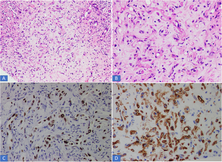
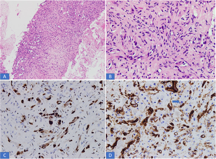
Results: Seven cases were reported during the study period. There were 4 females and 3 males. Age range was 20 to 77 years, mean age was 45 years. Three patients presented with right upper abdominal pain; 1 patient presented with jaundice while 3 patients were asymptomatic. In all 7 cases, lesions were identified on imaging studies. In 5 cases, liver lesions were multifocal. Clinical differential diagnosis in all cases was metastatic carcinoma and multifocal hepatocellular carcinoma. Liver function tests were normal in 5 cases. In 1 patient, tumor had already metastasized to the right lung. On histological examination of liver core biopsies performed in all 7 cases, classic histological features of epithelioid hemangioendothelioma were seen. Tumor cells expressed positivity for vascular markers (CD 34, CD31 and ERG) and were negative for cytokeratins, Hep par 1 and Glypican 3. Surgical resection was not performed in any of the 7 cases and all patients were treated by chemotherapy. Follow up was available in 5 cases. Of these, 3 patients died of disease and another patient was alive with metastases in both lungs, omentum and colon.
Conclusion: Clinicopathological features of the 7 cases in our series and detailed review of published literature is presented. Prognosis was bad in our cases most likely due to fact that surgical resection could not be performed in any of the cases owing to lack of surgical expertise for liver tumor surgery in most parts of the country.
Keywords: CD31; CD34; ERG; Epithelioid hemangioendothelioma; Hepatic; Multifocal.
Conflict of interest statement
The authors declare that they have no competing interests.
Figures

References
-
- Hornick JL. Epithelioid hemangioendothelioma. In WHO Classification of Tumors Editorial Board. WHO Classification of Tumors 5th Edition. Digestive System Tumors. Lyon: IARC 2019:466–7.
-
- Studer LL, Selby DM. Hepatic epithelioid hemangioendothelioma. Arch Pathol Lab Med. 2018;142:263–267. - PubMed
-
- Mehrabi A, Kashfi A, Fonouni H, Schemmer P, Halscheidt P, et al. Primary malignant hepatic epithelioid hemangioendothelioma. A comprehensive review of the literature with emphasis on the surgical therapy. Cancer. 2006;107(9):2108–2121. - PubMed
MeSH terms
Substances
LinkOut - more resources
Full Text Sources
Medical
Research Materials
Miscellaneous
