. 2020 Sep 25;6(39):eabc1726.
doi: 10.1126/sciadv.abc1726. Print 2020 Sep.

The prefusion structure of herpes simplex virus glycoprotein B

Affiliations

The prefusion structure of herpes simplex virus glycoprotein B

B Vollmer et al. Sci Adv. .

Abstract

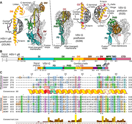
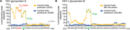
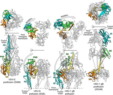
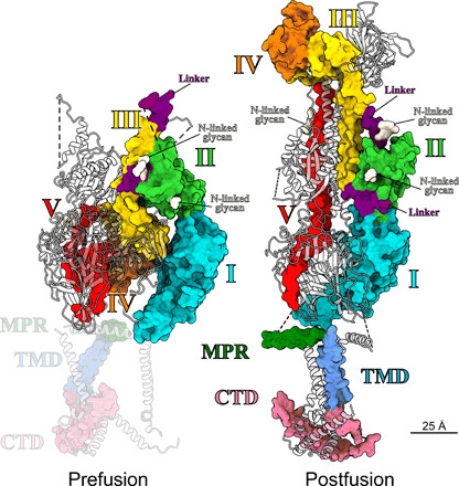
Cell entry of enveloped viruses requires specialized viral proteins that mediate fusion with the host membrane by substantial structural rearrangements from a metastable pre- to a stable postfusion conformation. This metastability renders the herpes simplex virus 1 (HSV-1) fusion glycoprotein B (gB) highly unstable such that it readily converts into the postfusion form, thereby precluding structural elucidation of the pharmacologically relevant prefusion conformation. By identification of conserved sequence signatures and molecular dynamics simulations, we devised a mutation that stabilized this form. Functionally locking gB allowed the structural determination of its membrane-embedded prefusion conformation at sub-nanometer resolution and enabled the unambiguous fit of all ectodomains. The resulting pseudo-atomic model reveals a notable conservation of conformational domain rearrangements during fusion between HSV-1 gB and the vesicular stomatitis virus glycoprotein G, despite their very distant phylogeny. In combination with our comparative sequence-structure analysis, these findings suggest common fusogenic domain rearrangements in all class III viral fusion proteins.


Figures

Fig. 1
Fig. 1. Determination of a conserved hinge in DIII of gB.
(A) Trimeric ectodomain x-ray structures of gB in postfusion (6) and VSV-G in postfusion (11) and prefusion (12) conformation. Corresponding domains are depicted using the same color scheme for a single protomer. The central helices of each of the protomers are magnified and displayed in insets. The hinge regions in gB RHV515–517 and VSV-G IQD272–274 are marked in striped green and the 310-helix VSL269–271 in red. Location of fusion loops is marked for one protomer. Domains are named according to (6) for gB and (16) for VSV-G. CD, central domain; PHD, pleckstrin homology domain; FD, fusion domain. (B) Domain architecture of gB and VSV-G. Numbers indicate amino acid positions of the domain boundaries. N-terminal signal peptides and the unstructured N-terminal domain of gB are shown in white, and flexible linker regions are shown in purple. CTD, C-terminal domain. (C) Sequence alignment of central helices of glycoprotein G [domain II (DII)] of Rhabdoviridae and glycoprotein B (DIII) of human Herpesviridae. The consensus secondary structure of the central postfusion coiled-coil helix of VSV (11) and HSV-1 (6) is shown in between the alignment of the two virus families. The color outlining the helix indicates the length of the helix of VSV-G (purple) and HSV-1 gB (orange), respectively, while the dashed outline marks the linker region to the C-terminally attached short helix. The residues forming the N-terminal 310-helix of the prefusion VSV-G structure are shown in red letters, and the hinge region residues of the same structure as well as the putative hinge in HSV-1 gB are shown in green letters. Residue background coloring is based on Clustal Omega (39), and conservation was calculated using www.compbio.dundee.ac.uk/aacon/ (40). Three highly conserved, sequence signatures are marked by asterisks on the bottom of the alignment. VHSV: viral hemorrhagic septicaemia virus; RABV: rabies virus; BEFV: bovine ephemeral fever virus; SIGV: Drosophila melanogaster sigma virus; VZV: varicella zoster virus; EBV: Epstein-Barr virus; HHV8: Kaposi’s sarcoma-associated herpesvirus; HHV6: human herpesvirus 6; HHV7: human herpesvirus 7; HCMV: human cytomegalovirus.
Fig. 2
Fig. 2. MD simulation of DIII in solution.
(A) Local bending angles of VSV-G DII in prefusion (yellow) (12) and postfusion (blue) (11) conformation. Protein Data Bank (PDB) numbers are given in parentheses. Amino acids are given in one-letter code. The 310-helix and hinge region are highlighted by dashed boxes in red and green, respectively. The α-helical part is highlighted by dashed box in gray. The position permitting a helix breaking mutation is marked with a black arrowhead. (B) Local bending angles of HSV-1 gB DIII in MD simulation (yellow) and postfusion (6) conformation (blue). The predicted hinge region in the MD simulation is marked in green. The position permitting a helix breaking mutation is marked with a black arrowhead.
Fig. 3
Fig. 3. Locking and functionally arresting the gB prefusion conformation.
(A) SDS–polyacrylamide gel electrophoresis (PAGE) and Coomassie stain of extracellular vesicle purifications, obtained from cells transfected with the HSV-1 gB wild type (wt) and single-point mutants, and Western blot analysis of the cell lysate and extracellular vesicles. Mature gB protein is marked by a black arrowhead. Used antibodies are indicated on the right. GAPDH: glyceraldehyde-3-phosphate dehydrogenase. (B) Fusion activity of gB wt, a fusion-null construct, or a single-point mutant in combination with gH/gL and gD in a cell-cell fusion assay. Activities are normalized to wt fusion activity level. (C) Cryo-ET slices of purified extracellular vesicles formed by WT and gB H516P. The long, postfusion form of gB is indicated by blue lines, and the short form is indicated by orange lines. Lower left images show the top views of gB trimers. Scale bars: 25 nm. (D) Size distribution of vesicles found with short and long gB form on vesicles formed by wt gB (n = 183) and gB H516P (n = 114). Boxes indicate range from first to third quartile with whiskers showing ±1.5 interquartile range and outliers marked as points. *no range given, as only two vesicles were found displaying the long form of gB H516P. The red line marks the median diameter.
Fig. 4
Fig. 4. Structure of the locked gB protein.
(A) Top and side view of the SVA structure, acquired by cryo-ET of gB H516P. Dashed lines mark the positions of the alphabetically marked cross sections on the right (a to h). White arrowheads point to the neighboring gB trimers. (B) Front slice of the fitted ectodomain structure showing the linker region connecting DII and DIII. Red arrowheads mark the β hairpin in DI protruding outside of the SVA map. Small insets at the top left indicate the direction of view and the section seen. Domains are marked and colored as in (6). (C) Middle slice showing the fit of DIII and DV in the central tubular densities. (D) Top view showing the crest region and fitted DII and DIII. (E) Bottom slice seen from above showing the fusion loops and central, tubular densities accommodating DV.
Fig. 5
Fig. 5. Comparison of the domain rearrangement between pre- and postfusion conformation in VSV-G and gB.
The structures of VSV-G (11, 12) and HSV-1 gB (6) are shown in side (lower row) and top (upper row) views in pipe representation with one protomer colored as in Fig. 1A. For clarity, DV of gB is omitted, and to increase visibility, one protomer is framed by a black silhouette. The prefusion conformation of each protein is depicted in the middle with the postfusion conformation on the sides. The pre- and postfusion conformation of each structure is aligned to the region including DIV (orange) and the C-terminal helix of DIII (yellow) in gB or the CD region in VSV-G, respectively. The one-sided gray arrows indicate the movement of DI (blue) and DII (green) in gB and the corresponding FD (blue) and PHD (green) in VSV-G in relation to the rest of the protein with the upward movement shown in the side views and the lateral movement shown in the top views. The hinge regions in DIII of gB and the CD in VSV-G are shown in striped green with the conserved sequence signatures marked by black asterisks.
Fig. 6
Fig. 6. Domain architecture of the pre- and postfusion conformation.
Structure of the rigid-body fit for the prefusion conformation in comparison to the full-length postfusion structure. Domains are marked and colored as in (14), also indicating representative positions of potential N-linked glycosylations. Two protomers per structure are shown as gray ribbons. For clarity, ribbons are half transparent. Because of the uncertainty of the fit, the MPR, TMD, and C-terminal domain (CTD) of the prefusion structure are shaded.

References

    1. Harrison S. C., Viral membrane fusion. Virology 479-480, 498–507 (2015). - PMC - PubMed
    1. McLellan J. S., Chen M., Leung S., Graepel K. W., Du X., Yang Y., Zhou T., Baxa U., Yasuda E., Beaumont T., Kumar A., Modjarrad K., Zheng Z., Zhao M., Xia N., Kwong P. D., Graham B. S., Structure of RSV fusion glycoprotein trimer bound to a prefusion-specific neutralizing antibody. Science 340, 1113–1117 (2013). - PMC - PubMed
    1. McLellan J. S., Chen M., Joyce M. G., Sastry M., Stewart-Jones G. B. E., Yang Y., Zhang B., Chen L., Srivatsan S., Zheng A., Zhou T., Graepel K. W., Kumar A., Moin S., Boyington J. C., Chuang G.-Y., Soto C., Baxa U., Bakker A. Q., Spits H., Beaumont T., Zheng Z., Xia N., Ko S.-Y., Todd J.-P., Rao S., Graham B. S., Kwong P. D., Structure-based design of a fusion glycoprotein vaccine for respiratory syncytial virus. Science 342, 592–598 (2013). - PMC - PubMed
    1. Krarup A., Truan D., Furmanova-Hollenstein P., Bogaert L., Bouchier P., Bisschop I. J. M., Widjojoatmodjo M. N., Zahn R., Schuitemaker H., McLellan J. S., Langedijk J. P. M., A highly stable prefusion RSV F vaccine derived from structural analysis of the fusion mechanism. Nat. Commun. 6, 8143 (2015). - PMC - PubMed
    1. M. Backovic, T. S. Jardetzky, in Cell Fusion in Health and Disease II: Cell Fusion in Disease (2011), vol. 714, pp. 91–101.

Publication types