. 2020 Sep 25;6(39):eaba9338.
doi: 10.1126/sciadv.aba9338. Print 2020 Sep.

A machine learning approach to define antimalarial drug action from heterogeneous cell-based screens

Affiliations

A machine learning approach to define antimalarial drug action from heterogeneous cell-based screens

George W Ashdown et al. Sci Adv. .

Abstract

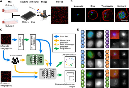
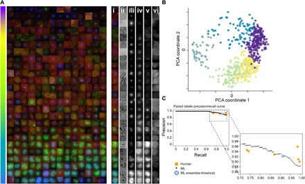
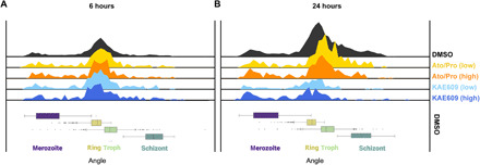
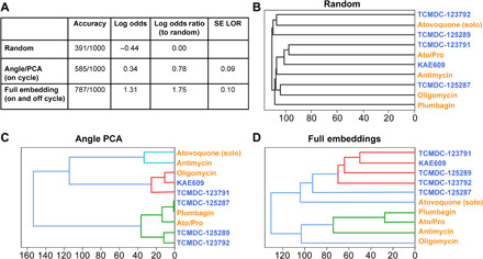
Drug resistance threatens the effective prevention and treatment of an ever-increasing range of human infections. This highlights an urgent need for new and improved drugs with novel mechanisms of action to avoid cross-resistance. Current cell-based drug screens are, however, restricted to binary live/dead readouts with no provision for mechanism of action prediction. Machine learning methods are increasingly being used to improve information extraction from imaging data. These methods, however, work poorly with heterogeneous cellular phenotypes and generally require time-consuming human-led training. We have developed a semi-supervised machine learning approach, combining human- and machine-labeled training data from mixed human malaria parasite cultures. Designed for high-throughput and high-resolution screening, our semi-supervised approach is robust to natural parasite morphological heterogeneity and correctly orders parasite developmental stages. Our approach also reproducibly detects and clusters drug-induced morphological outliers by mechanism of action, demonstrating the potential power of machine learning for accelerating cell-based drug discovery.


Figures

Fig. 1
Fig. 1. Experimental workflow.
(A) To ensure all life cycle stages were present during imaging and analysis, two transgenic malaria cultures, continuously expressing sfGFP, were combined (see Materials and Methods); these samples were incubated with or without drugs before being fixed and stained for automated multichannel high-resolution, high-throughput imaging. Resulting datasets (B) contained parasite nuclei (blue), cytoplasm (not shown), and mitochondrion (green) information, as well as the RBC plasma membrane (red) and brightfield (not shown). Here, canonical examples of a merozoite, ring, trophozoite, and schizont stage are shown. These images were processed for ML analysis (C) with parasites segregated from full field of views using the nuclear stain channel, before transformation into embedding vectors. Two networks were used; the first (green) was trained on canonical examples from human-labeled imaging data, providing ML–derived labels (pseudolabels) to the second semi-supervised network (gray), which predicted life cycle stage and compound phenotype. Example images from human-labeled datasets (D) show that disagreement can occur between human labelers when categorizing parasite stages (s, schizont; t, trophozoite; r, ring; m, merozoite). Each thumbnail image shows (from top left, clockwise) merged channels, nucleus staining, cytoplasm, and mitochondria. Scale bar, 5 μm.
Fig. 2
Fig. 2. ML continuum of parasite stages.
After learning from canonical human-labeled parasite images (for examples, please see Fig. 1B) and filtering debris and other outliers, the remaining life cycle data from asynchronous cultures was successfully ordered by the model. The parasites shown are randomly selected DMSO control parasites from multiple imaging runs, sorted by Angle PCA (A). The colored, merged images show RBC membrane (red), mitochondria (green), and nucleus (blue). For a subset of parasites on the right, the colored, merged image plus individual channels are shown: (i) merged, (ii) brightfield minimum projection, (iii) nucleus, (iv) cytoplasm, (v) mitochondria, and (vi) RBC membrane (brightfield maximum projection was also used in ML but is not shown here). The model sorts the parasites in life cycle stage order, despite heterogeneity of signal due to nuances such as imaging differences between batches. The order of the parasites within the continuum seen in (A) is calculated from the angle within the circle created by projecting model outputs using PCA, creating a 2D scatterplot (B). This represents a progression through the life cycle stages of the parasite, from individual merozoites (purple) to rings (yellow), trophozoites (green), schizonts (dark green), and finishing with a cluster of merozoites (blue). The precision-recall curve (C) shows that human labelers and the model have equivalent accuracy in determining the earlier/later parasite in pairs. The consensus of the human labelers was taken as ground truth, with individual labelers (orange) agreeing with the consensus on 89.5 to 95.8% of their answers. Sweeping through the range of “too close to call” values with the ML model yields the ML curve shown in black. Setting this threshold to 0.11 radians, the median angle variance across the individual models used in the ensemble yields the blue dot.
Fig. 3
Fig. 3. Quantifying on-cycle drug effects.
Asynchronous Plasmodium falciparum cultures were treated with the ATPase4 inhibitor KAE609 or the combinational MITO treatment of atovaquone and proguanil (Ato/Pro) with samples fixed and imaged 6 (A) and 24 (B) hours after drug additions. Top panels show histograms indicating the number of parasites across life cycle continuum. Compared to DMSO controls (topmost black histogram), both treatments demonstrated reduced parasite numbers after 24 hours. Shown are four drug/concentration treatment conditions: low-dose Ato/Pro (yellow), high-dose Ato/Pro (orange), low-dose KAE609 (light blue), and high-dose KAE609 (dark blue). Box plots below demonstrate life cycle classifications in the DMSO condition of images from merozoites (purple) to rings (yellow), trophozoites (green), and finishing with schizonts (dark green).
Fig. 4
Fig. 4. Quantifying off-cycle drug effects.
To better define drug effect on Plasmodium falciparum cultures, five mitochondrial (orange text) and five PfATP4ase (blue text) compounds were used; after a 24-hour incubation, images were collected and analyzed by the semi-supervised model. To test performance, various conditions were used (A). For random, images and drug names were scrambled, leading to the model incorrectly grouping compounds based on known MoA (B). Using life cycle stage definition (as with Fig. 3), the model generated improved grouping of compounds (C) versus random. Last, by combining the life cycle stage information with the penultimate layer (morphological information, before life cycle stage definition) of the model, it led to correct segregation of drugs based on their known MoA (D).

References

    1. Moffat J. G., Vincent F., Lee J. A., Eder J., Prunotto M., Opportunities and challenges in phenotypic drug discovery: An industry perspective. Nat. Rev. Drug Discov. 16, 531–543 (2017). - PubMed
    1. Antonova-Koch Y., Meister S., Abraham M., Luth M. R., Ottilie S., Lukens A. K., Sakata-Kato T., Vanaerschot M., Owen E., Jado J. C., Maher S. P., Calla J., Plouffe D., Zhong Y., Chen K., Chaumeau V., Conway A. J., McNamara C. W., Ibanez M., Gagaring K., Serrano F. N., Eribez K., Taggard C. M., Cheung A. L., Lincoln C., Ambachew B., Rouillier M., Siegel D., Nosten F., Kyle D. E., Gamo F.-J., Zhou Y., Llinás M., Fidock D. A., Wirth D. F., Burrows J., Campo B., Winzeler E. A., Open-source discovery of chemical leads for next-generation chemoprotective antimalarials. Science 362, eaat9446 (2018). - PMC - PubMed
    1. WHO, World Malaria Report (Geneva, 2019).
    1. Hamilton W. L., Amato R., van der Pluijm R. W., Jacob C. G., Quang H. H., Thuy-Nhien N. T., Hien T. T., Hongvanthong B., Chindavongsa K., Mayxay M., Huy R., Leang R., Huch C., Dysoley L., Amaratunga C., Suon S., Fairhurst R. M., Tripura R., Peto T. J., Sovann Y., Jittamala P., Hanboonkunupakarn B., Pukrittayakamee S., Chau N. H., Imwong M., Dhorda M., Vongpromek R., Chan X. H. S., Maude R. J., Pearson R. D., Nguyen T., Rockett K., Drury E., Gonçalves S., White N. J., Day N. P., Kwiatkowski D. P., Dondorp A. M., Miotto O., Evolution and expansion of multidrug-resistant malaria in southeast Asia: A genomic epidemiology study. Lancet Infect. Dis. 19, 943–951 (2019). - PMC - PubMed
    1. Wells T. N. C., Hooft van Huijsduijnen R., Van Voorhis W. C., Malaria medicines: A glass half full? Nat. Rev. Drug Discov. 14, 424–442 (2015). - PubMed

Publication types