. 2020 Dec 4;295(49):16700-16712.
doi: 10.1074/jbc.RA120.015360. Epub 2020 Sep 25.

Development and validation of a high-throughput whole cell assay to investigate Staphylococcus aureus adhesion to host ligands

Affiliations

Development and validation of a high-throughput whole cell assay to investigate Staphylococcus aureus adhesion to host ligands

Laurenne E Petrie et al. J Biol Chem. .

Abstract

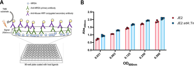
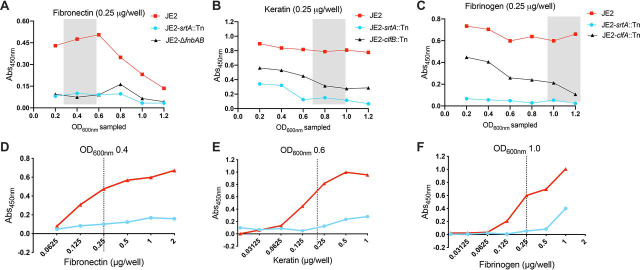
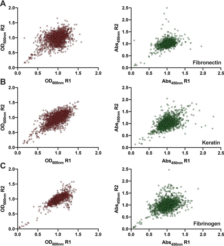
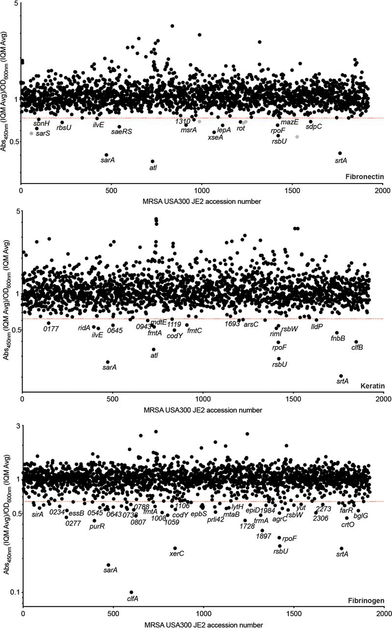
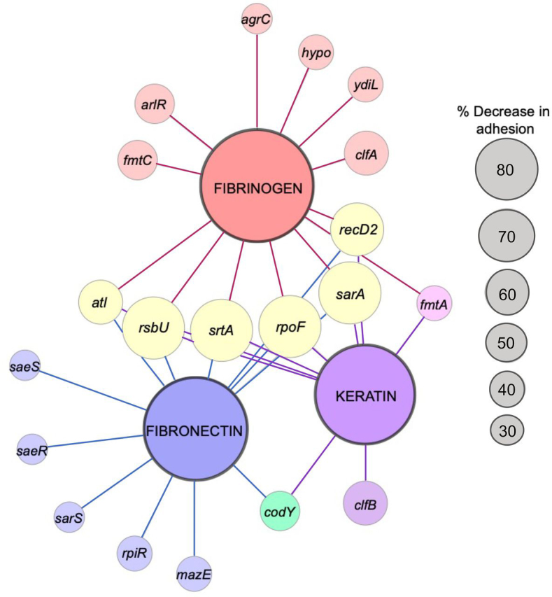
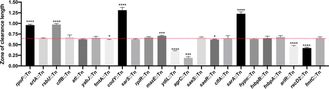
Staphylococcus aureus adhesion to the host's skin and mucosae enables asymptomatic colonization and the establishment of infection. This process is facilitated by cell wall-anchored adhesins that bind to host ligands. Therapeutics targeting this process could provide significant clinical benefits; however, the development of anti-adhesives requires an in-depth knowledge of adhesion-associated factors and an assay amenable to high-throughput applications. Here, we describe the development of a sensitive and robust whole cell assay to enable the large-scale profiling of S. aureus adhesion to host ligands. To validate the assay, and to gain insight into cellular factors contributing to adhesion, we profiled a sequence-defined S. aureus transposon mutant library, identifying mutants with attenuated adhesion to human-derived fibronectin, keratin, and fibrinogen. Our screening approach was validated by the identification of known adhesion-related proteins, such as the housekeeping sortase responsible for covalently linking adhesins to the cell wall. In addition, we also identified genetic loci that could represent undescribed anti-adhesive targets. To compare and contrast the genetic requirements of adhesion to each host ligand, we generated a S. aureus Genetic Adhesion Network, which identified a core gene set involved in adhesion to all three host ligands, and unique genetic signatures. In summary, this assay will enable high-throughput chemical screens to identify anti-adhesives and our findings provide insight into the target space of such an approach.

Keywords: MRSA; Staphylococcus aureus; anti-adhesives; antibiotic resistance; antivirulence; bacterial adhesion; cell wall-anchored proteins; drug discovery; high-throughput screening; methicillin-resistant Staphylococcus aureus (MRSA); virulence factor.


Conflict of interest statement

Conflict of interest—The authors declare that they have no conflicts of interest with the contents of this article.

Figures

Figure 1
Figure 1
An ELISA-based approach for the large-scale profiling of Staphylococcus aureus adhesion to host ligands.A, schematic diagram of the high-throughput S. aureus adhesion assay. B, recognition of surface-adhered MRSA USA300 by the primary antibody is not dependent on the presence of cell wall-anchored proteins. A Nunc™ MaxiSorp™ microtiter plate was coated with MRSA USA300 JE2 and isogenic JE2 srtA::Tn (devoid of CWAs) propagated to an OD600nm of 0.6. The cells were harvested, washed, and standardized to the various optical densities shown. Using the described ELISA, detection of surface adhered JE2 srtA::Tn was equivalent to that of the parental strain, indicating that recognition by the primary antibody is independent of CWA surface proteins. The values shown are the average mean ± S.D. of three duplicate biological replicates.
Figure 2
Figure 2
Optimization of the whole cell adhesion assay. The affinity of S. aureus for host ligands changes throughout the cell cycle; the strains were sampled at the different OD600nm shown and standardized to an OD600nm of 1.0. Adhesion to fibronectin (A), keratin (B), and fibrinogen (C), was assessed using the described ELISA (as shown in Fig. 1A), by measurement of the A450nm. For each host ligand, subsequent adhesion studies were performed with strains grown to the OD600nm range highlighted by the gray box. This range was selected based on the OD600nm where MRSA USA300 JE2 adhesion was maximal and presented an optimum ratio of separation between MRSA USA300 JE2 and the isogenic mutants of each respective ligand. To optimize the screening window, dose-dependent ELISAs were performed with MRSA USA300 JE2 and isogenic JE2 srtA::Tn adhering to human-derived fibronectin (D), keratin (E), and fibrinogen (F). The strains were grown to the specified OD600nm highlighted by the gray box and standardized to an OD600nm of 1.0. The dashed lines indicate the chosen concentration of each ligand for the subsequent transposon library adhesion screens. The values shown are the mean of three technical replicates.
Figure 3
Figure 3
Replica plots of normalized growth and adhesion values from the large-scale profiling of an MRSA USA300 transposon library (21). Growth (OD600nm) is shown on the left (red) and adhesion (A450nm) values are shown on the right (green). A, fibronectin; B, keratin; C, fibrinogen. R1 = replicate 1 and R2 = replicate 2.
Figure 4
Figure 4
Identifying genetic loci associated with S. aureus adhesion to human-derived fibronectin, keratin, and fibrinogen, respectively. The values shown in the index plots are the ratio between the average of duplicate adhesion values (A450nm) and duplicate growth values (OD600nm) (see Fig. 3). The strains are ordered based on their associated SAUSA300 accession number (21). The screens were performed with ligand concentrations and strains sampled at the given time points depicted in Fig. 2. Strains falling below the red lines exhibited a ratio less than 4 standard deviations (fibronectin and keratin) and 6 standard deviations (fibrinogen) from the IQM (75) of the data set.
Figure 5
Figure 5
The S. aureus Genetic Adhesion Network and anti-adhesive target space. The larger central nodes identify the host ligand in each screen (fibrinogen, fibronectin, and keratin). The smaller nodes radiating from these points indicate transposons inserted into genes that caused a significant reduction (Student's t test, p value ≤ 0.05; see Table 1 and Fig. S1) in adhesion to the host ligand they are linked. The node size for each gene reflects the percent adhesion compared with the parental strain (i.e. the larger the node the more pronounced the attenuated adhesion). The figure was generated using Cytoscape. Genes mazE (SAUSA300_2026), recD2 (SAUSA300_1576), rpiR (SAUSA300_2264), and ydiL (SAUSA300_1984) are names provided on the basis of their predicted structure and function. One gene, labeled hypo (hypothetical protein), is unannotated (SAUSA300_0602).
Figure 6
Figure 6
Assessing the level of proteolytic activity in the adhesion attenuated mutants. Protease activity was assessed using TSA containing 1.5% Difco Skim Milk. The zones of clearance were measured using ImageJ. Each strain was measured with at least three biological replicates. Welch's t test (*, p ≤ 0.05; **, p ≤ 0.01; ***, p ≤ 0.001; ****, p ≤ 0.0001) was used to compare the mean length for each mutant to the mean of the parental strain (red dashed line).

References

    1. Leonard A.C., Petrie L.E., Cox G. Bacterial anti-adhesives: inhibition of Staphylococcus aureus nasal colonization. ACS Infect. Dis. 2019;5:1668–1681. doi: 10.1021/acsinfecdis.9b00193. 31374164. - DOI - PubMed
    1. Johnny W.P. Bacterial pathogenesis. In: Barron S., editor. Medical Microbiology. 4 Ed. 1996. Galveston, TX.
    1. Ofek I., Hasty D.L., Sharon N. Anti-adhesion therapy of bacterial diseases: prospects and problems. FEMS Immunol. Med. Microbiol. 2003;38:181–191. doi: 10.1016/S0928-8244(03)00228-1. 14522453. - DOI - PubMed
    1. Pizarro-Cerdá J., Cossart P. Bacterial adhesion and entry into host cells. Cell. 2006;124:715–727. doi: 10.1016/j.cell.2006.02.012. 16497583. - DOI - PubMed
    1. Sattin S., Bernardi A. Glycoconjugates and glycomimetics as microbial anti-adhesives. Trends Biotechnol. 2016;34:483–495. doi: 10.1016/j.tibtech.2016.01.004. 26875976. - DOI - PubMed

Uncited reference

    1. Bae T., Glass E.M., Schneewind O., Missiakas D. Generating a collection of insertion mutations in the Staphylococcus aureus genome using bursa aurealis. Methods Mol. Biol. 2008;416:103–116. doi: 10.1007/978-1-59745-321-9_7. 18392963. - DOI - PubMed

Publication types

MeSH terms