. 2020 Dec 4;295(49):16713-16731.
doi: 10.1074/jbc.RA120.015468. Epub 2020 Sep 25.

PFN2 and NAA80 cooperate to efficiently acetylate the N-terminus of actin

Affiliations

PFN2 and NAA80 cooperate to efficiently acetylate the N-terminus of actin

Rasmus Ree et al. J Biol Chem. .

Abstract

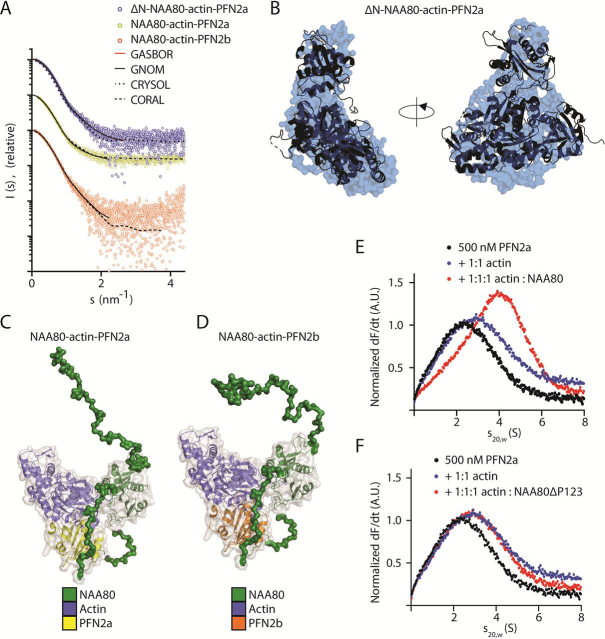
The actin cytoskeleton is of profound importance to cell shape, division, and intracellular force generation. Profilins bind to globular (G-)actin and regulate actin filament formation. Although profilins are well-established actin regulators, the distinct roles of the dominant profilin, profilin 1 (PFN1), versus the less abundant profilin 2 (PFN2) remain enigmatic. In this study, we use interaction proteomics to discover that PFN2 is an interaction partner of the actin N-terminal acetyltransferase NAA80, and further confirm this by analytical ultracentrifugation. Enzyme assays with NAA80 and different profilins demonstrate that PFN2 binding specifically increases the intrinsic catalytic activity of NAA80. NAA80 binds PFN2 through a proline-rich loop, deletion of which abrogates PFN2 binding. Small-angle X-ray scattering shows that NAA80, actin, and PFN2 form a ternary complex and that NAA80 has partly disordered regions in the N-terminus and the proline-rich loop, the latter of which is partly ordered upon PFN2 binding. Furthermore, binding of PFN2 to NAA80 via the proline-rich loop promotes binding between the globular domains of actin and NAA80, and thus acetylation of actin. However, the majority of cellular NAA80 is stably bound to PFN2 and not to actin, and we propose that this complex acetylates G-actin before it is incorporated into filaments. In conclusion, we reveal a functionally specific role of PFN2 as a stable interactor and regulator of the actin N-terminal acetyltransferase NAA80, and establish the modus operandi for NAA80-mediated actin N-terminal acetylation, a modification with a major impact on cytoskeletal dynamics.

Keywords: N-terminal acetyltransferases; NAT; acetylation; actin; cytoskeleton; post-translational modification (PTM); profilin; protein-protein interaction; small-angle X-ray scattering (SAXS).


Conflict of interest statement

Conflict of interest—The authors declare that they have no conflicts of interest with the contents of this article.

Figures

Figure 1
Figure 1
NAA80 co-immunoprecipitates PFN2.A, HeLa cells were transfected with NAA80-V5 or lacZ-V5 and B, HAP1 cells were transfected with V5-NAA80 or lacZ-V5. The V5 (IPs) were analyzed by LC–MS, and proteins were quantified by label-free quantification, comparing the intensity of each protein to the control IP (lacZ-V5) to identify interaction partners of NAA80. Red dots, significantly enriched proteins. Blue dots, selected nonenriched proteins. Dotted lines show log2 difference = 2 and -log10p value = 3.
Figure 2
Figure 2
NAA80 and PFN2 specifically interact, to the exclusion of PFN1.A–C, AUC experiments with fluorescently labeled profilin and varying ratios of unlabeled NAA80. D, V5 IPs in HAP1 cells (WT, PFN1-KO, or PFN2-KO) transfected with lacZ-V5 or V5-NAA80. Immunoprecipitates were probed with the indicated antibodies. Arrows indicate the expected sizes of the lacZ-V5, V5-NAA80, or actin bands. E, Western blotting-based absolute quantification of PFN1 and PFN2 in HAP1 cells, using purified proteins as standard. Right panel shows the quantification of the blots in the left panel (n = 3).
Figure 3
Figure 3
PFN2 increases the rate of actin acetylation in vitro.A, actin is fully acetylated in PFN1-KO and PFN2-KO cells. Western blots of lysates from the indicated cell types. B, schematic of the NAA80-profilin enzyme assays using β-actin N-terminal peptides (DDDIA) or full-length actin. C, NAA80/PFN DTNB enzyme assay using the N-terminal peptides of β-actin (n = 4). Enzyme reactions were run with NAA80/BSA for each ratio in parallel, and each NAA80/profilin ratio was normalized to the product formation for the corresponding NAA80/BSA ratio. Standard deviations were calculated as described under “Experimental procedures.” D, in vitro acetylation of full-length actin in the presence of PFN1 or PFN2b. Unacetylated actin purified from NAA80-KO cells was used as a substrate for NAA80/profilin. Right panel shows the quantification of the blots in the left panel (n = 3 for each condition). Error bars show S.D. Statistical significance was determined by Student's t test. n.s., p > 0.05; *, p < 0.05; **, p < 0.01.
Figure 4
Figure 4
The NAA80 polyproline-rich β6–β7 loop mediates PFN2 interaction.A, schematic of the NAA80 sequence, with secondary structure elements and regions of interest highlighted. Black, NAA80 isoform 1-specific N-terminus; yellow, Ac-CoA binding motif; red, polyproline stretches 1, 2, and 3. B, proline content in NAA80 sequences from the indicated species: Homo sapiens (Uniprot identifier: Q93015), Mus musculus (Q9R123), Rattus norvegicus (A0A0G2JV35), D. rerio (E7FBQ5), T. bimaculatus (A0A4Z2B4F3), Oryzias latipes (A0A3P9HTG7), P. trituberculatus (blue crab) (A0A5B7DRL4), Armadillidium vulgare (woodlouse) (A0A444SNE1), D. melanogaster (Q59DX8), A. gambiae (mosquito) (F5HLV5), and C. elegans (Q09518). Dashed line, average proline content in human genome (6.2%) (41). C, NAA80 β6–β7 loop alignment (residue range in brackets) of NAA80 from the same species as in B, aligned at β6 and β7, with prolines in red and gaps in gray. D, HAP1 cells were transfected with plasmids encoding V5-tagged NAA80-ΔP123 or lacZ/β-gal. V5-tagged proteins were immunoprecipitated and interactors were identified by LC–MS and LFQ, comparing the intensity of each protein to the control IP (lacZ-V5). Red dots, significantly enriched proteins. Blue dots, selected nonenriched proteins. Dotted lines show log2 difference = 2 and -log10p value = 3. E, heatmap of the LFQ intensities for the indicated protein groups in each IP. F and G, AUC with labeled PFN2a and NAA80 deletion mutants: NAA80-ΔP123 (F) or NAA80-ΔP1, NAA80-ΔP2, and NAA80-ΔP3 (G).
Figure 5
Figure 5
NAA80 requires PFN2 interaction for rapid actin Nt-acetylation.A, DTNB assay with NAA80-ΔP123 and varying molar ratios of PFN2a or PFN2b against DDDIA peptide (n = 3). Enzyme activity for each molar ratio was normalized to the activity of NAA80-ΔP123 alone. B, DTNB assay with NAA80-polyGS2 and varying molar ratios of PFN2a or PFN2b against DDDIA peptide (n = 3). Enzyme activity for each molar ratio was normalized to the activity of NAA80-polyGS2 alone. C, NAA80-KO cells were transfected with NAA80-WT or NAA80-ΔP123 for 8-14 h before Western blotting. Left, representative blots. Right, quantification of Ac-β- and Ac-γ-actin normalized to V5 expression from independent experiments (n = 4). *, p value <0.05, as determined by two-way ANOVA. See Fig. S7 for all quantified blots. Error bars show standard deviation.
Figure 6
Figure 6
NAA80 forms a ternary complex with PFN2 and actin.A, scattering profiles for ΔN-NAA80-actin-PFN2a, NAA80-actin-PFN2a, and NAA80-actin-PFN2b, with fits from GASBOR (red line), CORAL (dashed line), GNOM (solid line), and CRYSOL (dotted line). B, ab initio model of ΔN-NAA80-actin-PFN2a (surface representation), superimposed with the crystal structure of ΔN-NAA80-actin-PFN1 (PDB code 6NAS) in cartoon representation. C and D, SAXS models of NAA80-actin-PFN2a (C) or -PFN2b (D) in surface representation. The crystal structure of ΔN-NAA80-actin-PFN1 (cartoon representation) was used as rigid body, and the N-terminus and missing residues of the β6–β7 loop were modeled by using CORAL. E and F, analytical ultracentrifugation of labeled PFN2a, PFN2a with actin, and PFN2a with actin and full-length NAA80 (E) or NAA80-ΔP123 (F).
Figure 7
Figure 7
Gel filtration of cell lysates suggests the presence of low abundance NAA80-PFN2-actin complexes. Gel filtration chromatogram of HAP1 lysate and calibration mix using the same column and method. Fractions were analyzed by Western blotting and probed with the indicated antibodies. Arrows indicate sizes of standard proteins (670 kDa: bovine thyroglobulin; 150 kDa: bovine γ-globulins; 44.3 kDa: chicken egg albumin; 13.7 kDa: bovine pancreas RNase A). Double-headed arrow indicates specific NAA80 band (lower).
Figure 8
Figure 8
The function of NAA80 and PFN2 in actin N-terminal processing. During synthesis, NatB co-translationally acetylates the actin N-terminal and the acetylated initiator methionine (Ac-iMet) is subsequently removed by acetyl-methionine aminopeptidase (AcMetAP). The NAA80-PFN2 complex Nt-acetylates the actin neo-N-terminus and the Nt-acetylated actin subsequently enters the polymerization pool. Profilins PFN1/PFN2 bind G-actin with high affinity and promote binding of ATP and subsequent polymerization. ADP-actin dissociating at the pointed end binds profilin to reenter the cycle. Three possible options for how the newly synthesized actin enters the polymerization pool are presented: 1) after folding, actin is Nt-acetylated by NAA80-PFN2. NAA80-PFN2 facilitates ATP binding and delivers actin to the filament for polymerization, before being recycled for a new acetylation cycle. 2) After acetylation, NAA80 is released, PFN2 catalyzes actin-ATP binding, and actin is delivered to the filament. NAA80 binds PFN2 before a new acetylation cycle. 3) NAA80-PFN2 releases actin after acetylation and is immediately available for a new acetylation cycle. Actin is bound by profilin before ATP binding and delivery to the actin filament.

Similar articles

Cited by

References

    1. Pollard T.D., Cooper J.A. Actin, a central player in cell shape and movement. Science. 2009;326:1208–1212. doi: 10.1126/science.1175862. 19965462. - DOI - PMC - PubMed
    1. Varland S., Vandekerckhove J., Drazic A. Actin post-translational modifications: the cinderella of cytoskeletal control. Trends Biochem. Sci. 2019;44:502–516. doi: 10.1016/j.tibs.2018.11.010. 30611609. - DOI - PubMed
    1. Pollard T.D. Actin and actin-binding proteins. Cold Spring Harb. Perspect. Biol. 2016;8 doi: 10.1101/cshperspect.a018226. 26988969. - DOI - PMC - PubMed
    1. Courtemanche N., Pollard T.D. Interaction of profilin with the barbed end of actin filaments. Biochemistry. 2013;52:6456–6466. doi: 10.1021/bi400682n. 23947767. - DOI - PMC - PubMed
    1. Witke W. The role of profilin complexes in cell motility and other cellular processes. Trends Cell Biol. 2004;14:461–469. doi: 10.1016/j.tcb.2004.07.003. 15308213. - DOI - PubMed

Publication types

MeSH terms

LinkOut - more resources