. 2020 Dec 4;295(49):16732-16742.
doi: 10.1074/jbc.RA120.015694. Epub 2020 Sep 25.

α2-Macroglobulin-like protein 1 can conjugate and inhibit proteases through their hydroxyl groups, because of an enhanced reactivity of its thiol ester

Affiliations

α2-Macroglobulin-like protein 1 can conjugate and inhibit proteases through their hydroxyl groups, because of an enhanced reactivity of its thiol ester

Seandean Lykke Harwood et al. J Biol Chem. .

Erratum in

Abstract

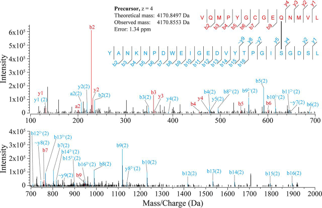
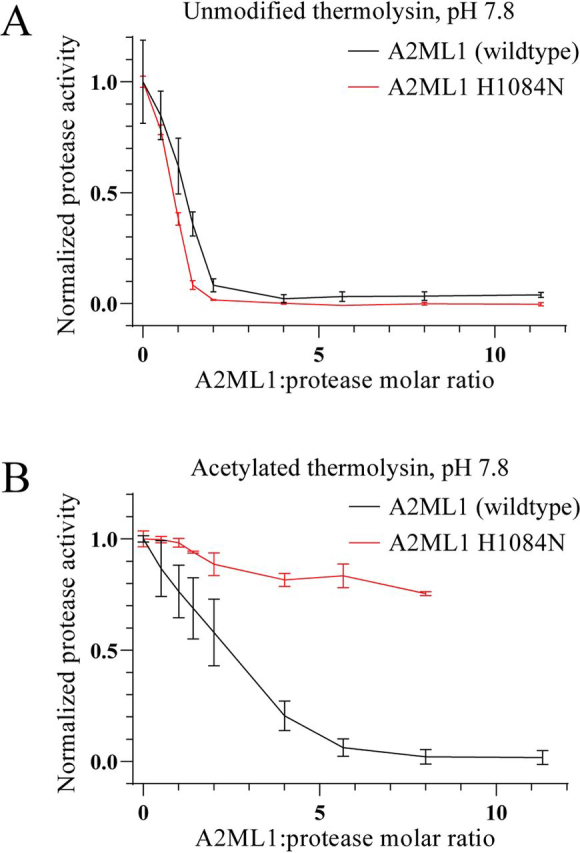
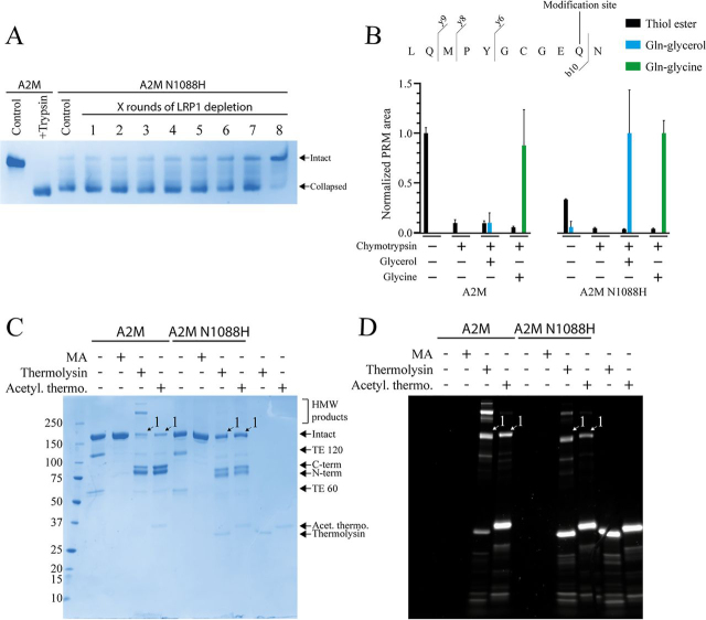
Proteins in the α-macroglobulin (αM) superfamily use thiol esters to form covalent conjugation products upon their proteolytic activation. αM protease inhibitors use theirs to conjugate proteases and preferentially react with primary amines (e.g. on lysine side chains), whereas those of αM complement components C3 and C4B have an increased hydroxyl reactivity that is conveyed by a conserved histidine residue and allows conjugation to cell surface glycans. Human α2-macroglobulin-like protein 1 (A2ML1) is a monomeric protease inhibitor but has the hydroxyl reactivity-conveying histidine residue. Here, we have investigated the role of hydroxyl reactivity in a protease inhibitor by comparing recombinant WT A2ML1 and the A2ML1 H1084N mutant in which this histidine is removed. Both of A2ML1s' thiol esters were reactive toward the amine substrate glycine, but only WT A2ML1 reacted with the hydroxyl substrate glycerol, demonstrating that His-1084 increases the hydroxyl reactivity of A2ML1's thiol ester. Although both A2ML1s conjugated and inhibited thermolysin, His-1084 was required for the conjugation and inhibition of acetylated thermolysin, which lacks primary amines. Using MS, we identified an ester bond formed between a thermolysin serine residue and the A2ML1 thiol ester. These results demonstrate that a histidine-enhanced hydroxyl reactivity can contribute to protease inhibition by an αM protein. His-1084 did not improve A2ML1's protease inhibition at pH 5, indicating that A2ML1's hydroxyl reactivity is not an adaption to its acidic epidermal environment.

Keywords: A2ML1; alpha 2 macroglobulin like protein 1; alpha-2-macroglobulin; complement; inhibition mechanism; mutagenesis; mutagenesis in vitro; protease; protease inhibitor; protein crosslinking; thiol ester; α2-macroglobulin.


Conflict of interest statement

Conflict of interest—The authors declare that they have no conflicts of interest with the contents of this article.

Figures

Figure 1
Figure 1
Reactivity of A2ML1's thiol ester toward amine and hydroxyl substrates.A, sequence alignment of A2ML1 and four other αM proteins showing the hydroxyl reactivity–conveying histidine possessed by A2ML1, C3, and C4B. B, WT and H1084N A2ML1 were cleaved by chymotrypsin in HBS with 200 mm of glycerol or glycine. The samples were acidified with formic acid to pH 3, digested using pepsin, and analyzed by LC-MS/MS. The major peptide covering the thiol ester site, with either an intact thiol ester or after conjugation to glycerol or glycine, was quantified by PRM. The fragmentation sites that produce the b10, b11, and b12 product ions used for quantification are indicated. C, PRM was used to quantify the peptide covering the thiol ester site with the three modifications. The sums of the b10, b11, and b12 product ion areas of each modified peptide were normalized to the sample in which that peptide was most abundant. Both A2ML1s were able to conjugate glycine, but formation of the glycerol reaction product was only detected using WT A2ML1. Quantification is based on three separately prepared samples per condition, and the error bars indicate the S.D. D, overlaid MS2 spectra from the three peptides. The b10, b11, and b12 product ions that were used for PRM quantification from each spectrum are indicated. As these product ions include the glutamine that participates in the thiol ester (Gln-973), their mass is increased by the mass of a glycerol or glycine after conjugation. The full annotated MS2 spectra for each peptide are shown in Fig. S1.
Figure 2
Figure 2
Reducing SDS-PAGE of A2ML1 digested with thermolysin.AD, WT (A and B) and H1084N (C and D) A2ML1 were digested with unmodified (A and C) or acetylated (B and D) thermolysin at various molar ratios of thermolysin:A2ML1, as indicated, for 15 min at 37°C, after which thermolysin was inhibited with 10 mm EDTA. Digestions were also performed with 50 mm BAPN present, which competes with proteases for thiol ester–mediated conjugation. An intact A2ML1 protein migrates as an ∼180-kDa band. When an intact thiol ester is present, some A2ML1 is fragmented when heated under denaturing conditions to ∼130-kDa and ∼50-kDa bands. Bait region cleavage gives an N-terminal cleavage product of ∼95 kDa and a C-terminal cleavage product of ∼85 kDa. The C-terminal cleavage product includes the thiol ester site and participates in protease conjugation products, which for thermolysin are ∼150 kDa and are indicated as band 1. This conjugation product can be further cleaved by thermolysin to yield band 2. In-gel digestion and MS were used to verify that bands 1 and 2 contain both A2ML1 and thermolysin. A third band, band 3, contains both the N-terminal and C-terminal fragments of A2ML1, but not thermolysin, and is because of intra-A2ML1 thiol ester conjugation, as shown in Fig. S4. Both A2ML1s are initially cleaved by thermolysin in their bait regions, with secondary cleavage elsewhere occurring at higher thermolysin ratios. Both A2ML1s form conjugation products to unmodified thermolysin, which has both hydroxyl and amine groups, but only WT A2ML1 conjugates to acetylated thermolysin, which only has hydroxyl groups. Digestion proceeds more readily in circumstances when protease conjugation is not possible, e.g. because of BAPN or when A2ML1 H1084N is cleaved by acetylated thermolysin.
Figure 3
Figure 3
An MS2 spectrum identifying conjugation of A2ML1's thiol ester to a thermolysin serine side chain. The SDS-PAGE band of the conjugation product between unmodified thermolysin and the C-terminal fragment of bait region–cleaved WT A2ML1, i.e. band 1 in Fig. 2, was digested with pepsin, extracted from the gel band, and analyzed by LC-MS/MS. Crosslinks between the thiol ester–covering peptide (VQMPYGCGEQNMVL) and thermolysin peptides were identified. In the above MS2 spectrum, HCD-fragmented products from the thiol ester peptide (in red) and a thermolysin peptide (in blue) cover both ends of each peptide and an extensive b product ion series (b4–b16) shows the lysine residue on the thermolysin peptide, K4, as unmodified. Fig. S3 conclusively demonstrates that K4 is not the site of conjugation, as the same experiment using acetylated thermolysin identified a crosslinked peptide identical to this one, except for acetylation of K4. Note that the ∼y7, ∼y8, and ∼y9 fragments show the putative site of conjugation S20 as unmodified. The other hydroxyl residues Y1, Y15, T16, and S23 are seen unmodified in fragments b2–b16, b15, b15/b16/y9, and y1/y2, respectively, and there are therefore no conjugation site candidate positions that are not seen as unmodified residues. However, the y82+ residue shows S20 crosslinked to the thiol ester peptide and thus supports S20 as the conjugation site. The spectra of additional crosslinked peptides are given in Fig. S3.
Figure 4
Figure 4
Inhibition of thermolysin by A2ML1.A and B, unmodified (A) and acetylated (B) thermolysin were incubated with the indicated ratios of A2ML1 or A2ML1 H1084N for 15 min at 37°C in 150 mm Tris-HCl, 15 mm CaCl2, pH 7.8. The samples were then incubated for an additional 60 min at 37°C with resorufin-labeled casein, after which TCA was added to terminate the reactions and precipitate intact substrate. Precipitated substrate was removed by filtration. The filtrate was neutralized and the residual proteolytic activity was determined by measuring the absorbance at 574 nm. n = 3 and the error bars show the S.D. Both A2ML1s inhibited unmodified thermolysin, requiring roughly a 2-fold excess of A2ML1 for full inhibition. WT A2ML1 inhibited acetylated thermolysin, albeit less efficiently, requiring a roughly 6-fold excess of A2ML1 for full inhibition, whereas A2ML1 H1084N demonstrated little to no inhibition.
Figure 5
Figure 5
SDS-PAGE analysis of A2ML1's conjugation to Cy5-labeled thermolysin at pH 5–7.AD, WT (A and B) and H1084N (C and D) A2ML1 were digested using a 1:1 molar ratio of thermolysin that was either only Cy5-labeled or was additionally acetylated after Cy5 labeling. Digestion was performed at pH 5, 6, or 7. Fluorescence images from 20 s of exposure visualizing the bands containing thermolysin (B and D) and images of the gels after Coomassie Blue staining (A and C) are shown. The major conjugation product between thermolysin and A2ML1 is labeled band 1, further processing of this product gives band 2, and intra-A2ML1 conjugation gives band 3, using the same numbering as in Fig. 2. Although both A2ML1s conjugate thermolysin similarly at pH 6 and 7, the ∼150 kDa major conjugation product (band 1) is moderately more intense at pH 5 for WT A2ML1 than for A2ML1 H1084N. Furthermore, acetylated thermolysin, which is only conjugated by WT A2ML1, yields a similarly intense conjugation product at pH 5 to those at pH 6 and 7, although it should be noted that secondary cleavage of the major conjugation product (e.g. yielding band 2) occurs to a lesser degree at pH 5 because of the lower activity of thermolysin at this pH, and the amount of ∼150 kDa conjugation is therefore not solely indicative of the degree to which conjugation takes place.
Figure 6
Figure 6
Thiol ester reactivity of A2M N1088H. The mutant A2M N1088H, in which the histidine conveying hydroxyl reactivity A2ML1 is introduced, was recombinantly expressed and characterized. A, pore-limited native PAGE of A2M N1088H before and after up to eight rounds of depletion using LRP1-loaded resin. Plasma-purified A2M in its intact native conformation and trypsin-cleaved collapsed conformation is included for comparison. A2M N1088H was expressed mostly in a collapsed conformation. However, the collapsed A2M N1088H could be depleted by using its binding to LRP1, allowing native A2M N1088H to be enriched. B, the reactivity of the plasma-purified and N1088H A2M thiol esters toward glycerol and glycine, as determined using MS PRM. The same approach used for A2ML1, as described in Fig. 1, was used for A2M. PRM quantification was based on the b10, y6, y8, and y9 fragments that cover Gln-975, which participates in the thiol ester and becomes conjugated to glycerol or glycine. Both A2Ms reacted similarly with the amine substrate glycine, but A2M N1088H showed an increased reactivity toward the hydroxyl substrate glycerol. Quantification is based on three separately prepared samples per condition, and the error bars indicate the S.D. C and D, reducing SDS-PAGE of plasma-purified and N1088H A2M treated with methylamine or cleaved with thermolysin that was either only Cy5-labeled or was additionally acetylated after Cy5 labeling. The Coomassie Blue–stained gel (C) and Cy5 fluorescence image (D) are shown. The conjugation product between the C-terminal bait region cleaved fragment of A2M and thermolysin is indicated as band 1, although this band co-migrates with that of the intact A2M subunit. Both A2Ms conjugated thermolysin and, to a lesser extent, acetylated thermolysin. A2M N1088H did not demonstrate an increased relative conjugation to acetylated thermolysin as compared with plasma-purified A2M.

Similar articles

Cited by

References

    1. Galliano M.F., Toulza E., Gallinaro H., Jonca N., Ishida-Yamamoto A., Serre G., Guerrin M. A novel protease inhibitor of the α2-macroglobulin family expressed in the human epidermis. J. Biol. Chem. 2006;281:5780–5789. doi: 10.1074/jbc.M508017200. 16298998. - DOI - PubMed
    1. Numata S., Teye K., Tsuruta D., Sogame R., Ishii N., Koga H., Natsuaki Y., Tsuchisaka A., Hamada T., Karashima T., Nakama T., Furumura M., Ohata C., Kawakami T., Schepens I. Anti-alpha-2-macroglobulin-like-1 autoantibodies are detected frequently and may be pathogenic in paraneoplastic pemphigus. J. Invest. Dermatol. 2013;133:1785–1793. doi: 10.1038/jid.2013.65. 23407400. - DOI - PubMed
    1. Larson E.D., Magno J.P.M., Steritz M.J., Llanes E., Cardwell J., Pedro M., Roberts T.B., Einarsdottir E., Rosanes R.A.Q., Greenlee C., Santos R.A.P., Yousaf A., Streubel S.O., Santos A.T.R., Ruiz A.G. A2ML1 and otitis media: Novel variants, differential expression, and relevant pathways. Hum. Mutat. 2019;40:1156–1171. doi: 10.1002/humu.23769. 31009165. - DOI - PMC - PubMed
    1. Santos-Cortez R.L., Chiong C.M., Reyes-Quintos M.R., Tantoco M.L., Wang X., Acharya A., Abbe I., Giese A.P., Smith J.D., Allen E.K., Li B., Cutiongco-de la Paz E.M., Garcia M.C., Llanes E.G., Labra P.J. Rare A2ML1 variants confer susceptibility to otitis media. Nat. Genet. 2015;47:917–920. doi: 10.1038/ng.3347. 26121085. - DOI - PMC - PubMed
    1. Caubet C., Jonca N., Brattsand M., Guerrin M., Bernard D., Schmidt R., Egelrud T., Simon M., Serre G. Degradation of corneodesmosome proteins by two serine proteases of the kallikrein family, SCTE/KLK5/hK5 and SCCE/KLK7/hK7. J. Invest. Dermatol. 2004;122:1235–1244. doi: 10.1111/j.0022-202X.2004.22512.x. 15140227. - DOI - PubMed

Publication types

MeSH terms