. 2020 Nov 5;10(11):4249-4269.
doi: 10.1534/g3.120.401386.

Identifying Key Genetic Regions for Cell Sheet Morphogenesis on Chromosome 2L Using a Drosophila Deficiency Screen in Dorsal Closure

Affiliations

Identifying Key Genetic Regions for Cell Sheet Morphogenesis on Chromosome 2L Using a Drosophila Deficiency Screen in Dorsal Closure

Stephanie M Fogerson et al. G3 (Bethesda). .

Abstract

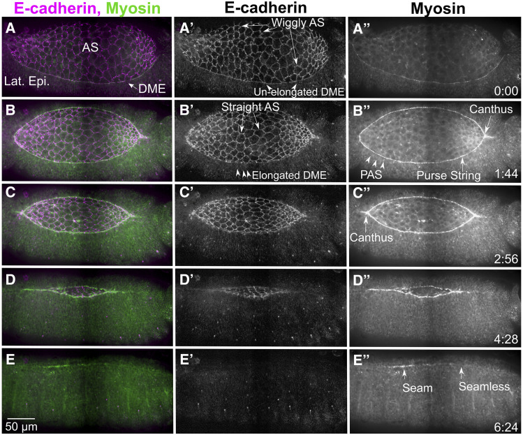
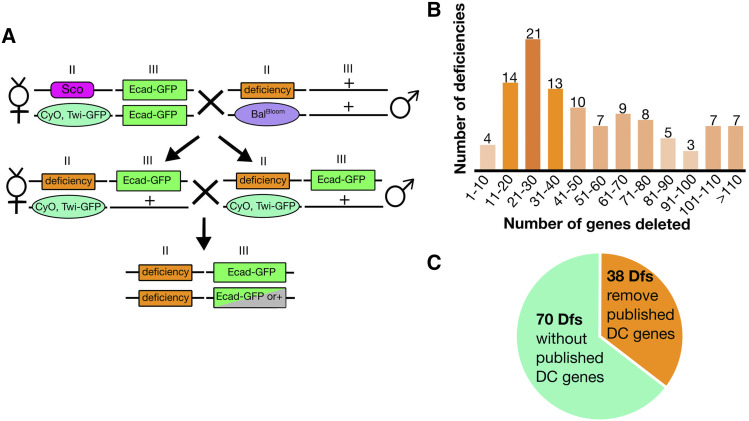
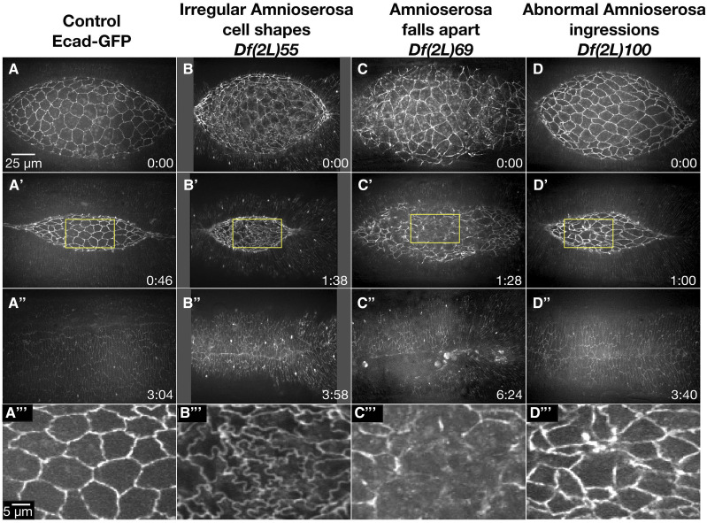
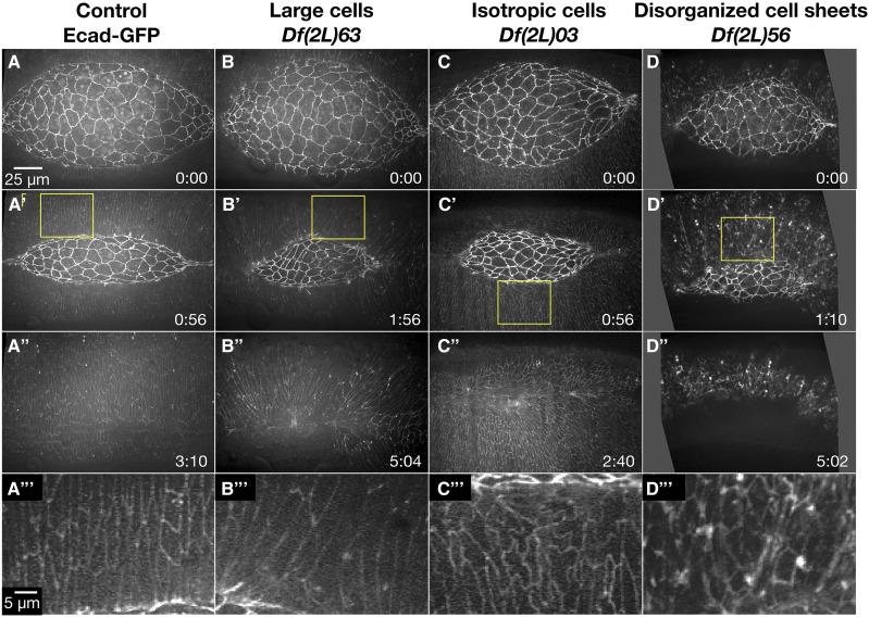
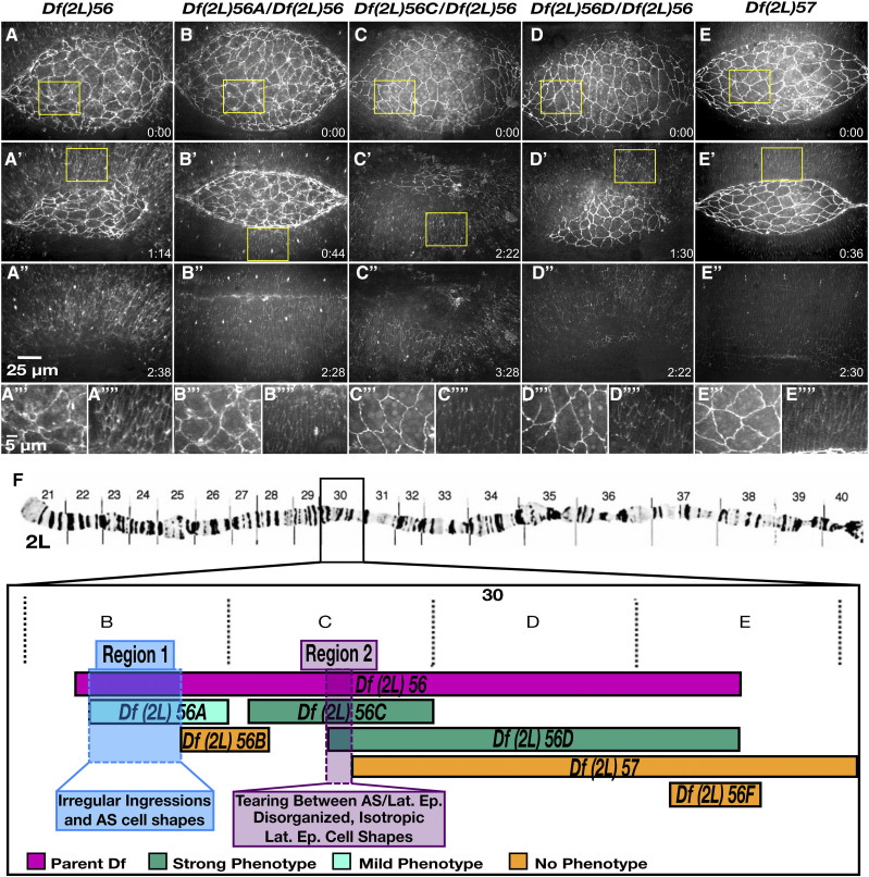
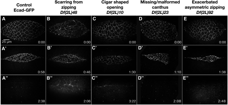
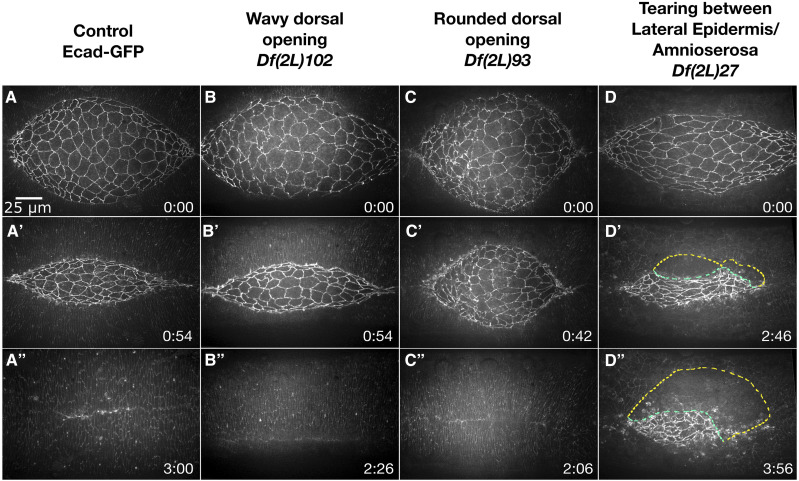
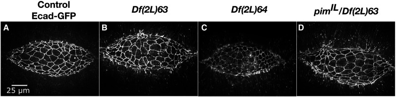
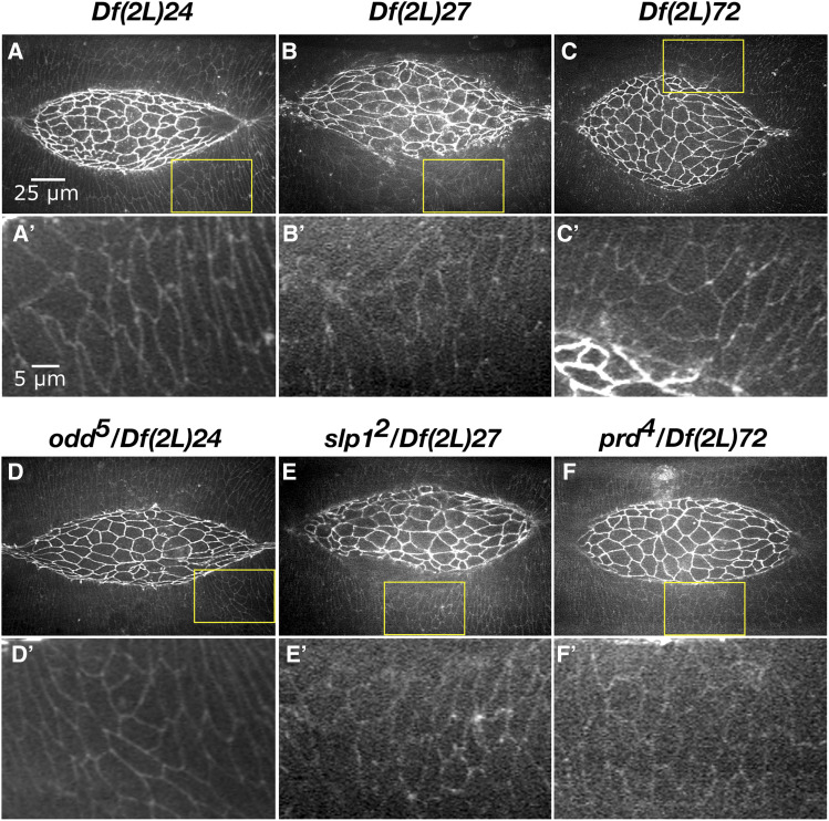
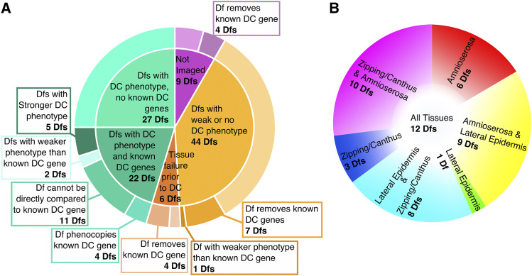
Cell sheet morphogenesis is essential for metazoan development and homeostasis of animal form - it contributes to developmental milestones including gastrulation, neural tube closure, heart and palate formation and to tissue maintenance during wound healing. Dorsal closure, a well-characterized stage in Drosophila embryogenesis and a model for cell sheet morphogenesis, is a remarkably robust process during which coordination of conserved gene expression patterns and signaling cascades regulate the cellular shape changes and movements. New 'dorsal closure genes' continue to be discovered due to advances in imaging and genetics. Here, we extend our previous study of the right arm of the 2nd chromosome to the left arm of the 2nd chromosome using the Bloomington deficiency kit's set of large deletions, which collectively remove 98.9% of the genes on the left arm of chromosome two (2L) to identify 'dorsal closure deficiencies'. We successfully screened 87.2% of the genes and identified diverse dorsal closure defects in embryos homozygous for 49 deficiencies, 27 of which delete no known dorsal closure gene. These homozygous deficiencies cause defects in cell shape, canthus formation and tissue dynamics. Within these deficiencies, we have identified pimples, odd-skipped, paired, and sloppy-paired 1 as dorsal closure genes on 2L that affect lateral epidermal cells. We will continue to identify novel 'dorsal closure genes' with further analysis. These forward genetic screens are expected to identify new processes and pathways that contribute to closure and links between pathways and structures already known to coordinate various aspects of closure.

Keywords: amnioserosa; dorsal closure; lateral epidermis; morphogenesis.


Figures

Figure 1
Figure 1
Dorsal closure progression from pre-canthus formation to a seamed epithelium. The cellular morphologies and cytoskeletal dynamics during dorsal closure are shown here by endogenously labeling cadherin at the adherens junctions (Ecad-Tomato, A’-E’) and myosin (myosin heavy chain-GFP exon trap, A”-E”) in stills taken from a stitched confocal time-lapse sequence. Prior to dorsal closure, the ends of the dorsal opening are blunt or rounded, the dorsal most epithelial (DME) cells are isotropic (unstretched), the amnioserosa have wiggly cell junctions and myosin is weakly localized to the boundary between the amnioserosa (AS) and lateral epidermis (Lat. Epi., A-A”) where the purse string will form. At the onset of dorsal closure, a canthus forms at the posterior end of the dorsal opening as zipping begins while the anterior end remains rounded (B-B”). The DME cells begin to elongate along their circumferential, dorsal-ventral axis (B’), while the peripheral amnioserosa (PAS) cells tuck under the DME cells (B”). The junctions of the bulk amnioserosa cells straighten and myosin bars accumulate at the purse string (B-B”). The anterior canthus soon forms (C-C”) and the lateral epidermal sheets zip together from both ends causing the dorsal opening to decrease in height (along the dorsal-ventral axis) and width (anterior-posterior axis, C-D”). Once dorsal closure completes, there is a seamed, and later seamless, epithelium (E-E”). Anterior is to the left, posterior to the right in all panels. Time is in hr:min. The scale bar in E applies to all panels (50 µm). Stills are from Supplemental Movie (Suppl Mov) 1, available on figshare.
Figure 2
Figure 2
Crossing scheme for live imaging deficiencies. Following two crosses, embryos homozygous for a Df can be selected by the lack of Twi-GFP and imaged with Ecad-GFP (A). The 2nd and 3rd chromosomes are indicated by Roman numerals II and III, respectively. The balancer from the Df stock is indicated by BalBloom. 2L Dfs remove as few as five genes and as many as 217 genes, the distribution of 2L Dfs by number of genes removed are shown, where the lightest orange represents 1-5 Dfs and the darkest orange represents 20 or more Dfs (B). Thirty-eight of the 108 2L Dfs remove genes documented to cause dorsal closure defects when mutated, while 70 Dfs do not (C).
Figure 3
Figure 3
Amnioserosa phenotypes observed in homozygous 2L Df embryos. Stills from a time-lapse sequence of Ecad-GFP labeled control embryos (A-A’’’) and examples of homozygous Df embryos that display amnioserosa phenotypes. Df(2L)55 embryos have irregular amnioserosa cell shapes (B-B’’’). In Df(2L)69 embryos, the amnioserosa cell sheet falls apart (C-C’’’). Df(2L)100 embryos show abnormal amnioserosa cell ingressions (D-D’’’). The yellow boxed areas in A’, B’, C’, and D’ are magnified in A’’’, B’’’, C’’’, and D’’’. Anterior is to the left, posterior to the right. Time is in hr:min, time 0:00 is at the start of the experimental run when the height of the dorsal opening was between 75-100 µm. The scale bar in A applies to panels A-D” and the scale bar in A’’’ applies to panels A’’’-D’’’. Stills are from Suppl Movies 2-5.
Figure 4
Figure 4
Lateral epidermal phenotypes observed in homozygous 2L Df embryos. Stills from a time-lapse sequence of Ecad-GFP labeled control embryos (A-A’’’) and homozygous Df embryos with lateral epidermal phenotypes. Homozygous Df(2L)63 embryos have large lateral epidermal cells (B-B’’’). Homozygous Df(2L)03 embryos have isotropic lateral epidermal cells (C-C’’’). Homozygous Df(2L)56 embryos have disorganized lateral epidermal cell sheets (D-D’’’). The yellow boxed areas in A’, B’, C’, and D’ are magnified in A’’’, B’’’, C’’’, and D’’’. Anterior is to the left, posterior to right. Time is in hr:min, time 0:00 is at the start of the experimental run when the height of the dorsal opening was between 75-100 µm. The scale bar in A applies to panels A-D”. The scale bar in A’’’ applies to panels A’’’-D’’’. Stills are from Suppl Movies 6-9, Supp Movies 10 and 24 provide additional examples of Df(2L)56 and Df(2L)63, respectively.
Figure 5
Figure 5
The severe and complex phenotype in embryos homozygous for Df(2L)56 can be divided into three separate phenotypes due to three distinct genomic regions. Stills from time-lapse sequence of homozygous Df(2L)56 (A-A’’’’), transheterozygous Df(2L)56A / Df(2L)56 (B-B’’’’), transheterozygous Df(2L)56C / Df(2L)56 (C-C’’’’), transheterozygous Df(2L)56D / Df(2L)56 (D-D’’’’) and homozygous Df(2L)57 embryos (E-E’’’’) in a DE-cadherin-GFP imaging background. Homozygous embryos of Df(2L)56 have a strong and fails dorsal closure phenotype. Transheterozygous embryos of Df(2L)56 with Df(2L)56A have a mid-severity dorsal closure phenotype with irregular amnioserosa cell shapes. Embryos transheterozygous for Df(2L)56C / Df(2L)56 as well as embryos transheterozygous for Df(2L)56D / Df(2L)56 have a ‘strong and fails’ phenotype –the amnioserosa tears away from the lateral epidermis and the lateral epidermal cell sheet has disorganized, isotropic cell shapes. Homozygous embryos of Df(2L)57 have no dorsal closure phenotype. The yellow boxed areas in A, A’, B, B’, C, C’, D, D’, E and E’ are magnified in A’’’, A’’’’, B’’’, B’’’’, C’’’, C’’’’, D’’’, D’’’’, E’’’ and E’’’’, respectively. A cytological map schematic of the left arm of chromosome 2 depicts the region removed in Df(2L)56 and overlapping sub-Dfs (F). The polytene chromosome micrograph was previously published in Halsell and Kiehart (1998) and Lefevre (1976). Embryos transheterozygous for Df(2L)56F / Df(2L)56 have no dorsal closure phenotypes (denoted in orange). Anterior is to the left, posterior to right. Time is in hr:min, time 0:00 is at the start of the experimental run when the height of the dorsal opening was between 75-100 µm. The scale bar in A applies to panels A-D’’ and the scale bar in A’’’ applies to A’’’-E’’’’. Stills are from Suppl Movies 10-14, Supp Mov 9 provides an additional example of Df(2L)56.
Figure 6
Figure 6
Zipping and canthus phenotypes observed in homozygous 2L Df embryos. Stills from time-lapse sequences of Ecad-GFP labeled control embryos (A-A”) and homozygous Df embryos with zipping and or canthus defects. Df(2L)48 embryos show scarring from zipping (B-B’’). Df(2L)10 embryos have a cigar-shaped dorsal opening (C-C’’). Df(2L)23 embryos are missing the posterior canthus (D-D’’). Df(2L)92 embryos have exacerbated asymmetric zipping: the anterior canthus zips faster than the posterior (E-E’’). Note that the timepoint was chosen as it illustrates the extreme asymmetry which is driven by increased ingression; however, a smaller more apical z-projection would show seam formation, but doesn’t illustrate zipping as well. Anterior is to the left, posterior to the right. Time is in hr:min, time 0:00 is at the start of the experimental run when the height of the dorsal opening was between 75-100 µm. The scale bar in A applies to all micrographs. Stills are from Suppl Movies 15-19.
Figure 7
Figure 7
Phenotypes at the interface of the peripheral amnioserosa and dorsal-most epithelium observed in homozygous 2L Df embryos. Stills from a time-lapse sequence of Ecad-GFP labeled control embryos (A-A”) and homozygous Df embryos with phenotypes at the interface of the peripheral amnioserosa and dorsal-most epithelium. A wavy dorsal opening is observed in Df(2L)102 embryos (B-B’’). Df(2L)93 embryos have a rounded dorsal opening (C-C’’). The peripheral amnioserosa tears away from the dorsal-most epithelium in Df(2L)27 embryos (D-D’’). The yellow dashed line mark the border of the lateral epidermis and the green dashed line marks the amnioserosa edge. Anterior is to the left, posterior to right. Time is in hr:min, time 0:00 is at the start of the experimental run when the height of the dorsal opening was between 75-100 µm. The scale bar in A applies to all micrographs. Stills are from Suppl Movies 20-23, note that Supp Mov 30 provides an additional example of Df(2L)27.
Figure 8
Figure 8
Deletion of pim is responsible for the large lateral epidermal cells of homozygous Df(2L)63 and Df(2L)64 embryos. Stills from a time-lapse sequence of mid-dorsal closure stage, Ecad-GFP labeled control (A), homozygous Df(2L)63 (B), Df(2L)64 (C) and transheterozygous pimIL / Df(2L)63 (D) embryos. Anterior is to the left, posterior to right. The scale bar in A applies to all micrographs. Stills are from Suppl Movies 24-27, Supp Mov 7 provides an additional example of Df(2L)63.
Figure 9
Figure 9
Pair-rule genes and the Dfs that remove them cause reduced elongation of lateral epidermal cells toward the dorsal midline (circumferentially along the dorsal-ventral axis). Stills from a time-lapse sequences of mid-dorsal closure stage, homozygous Df(2L)24 (A), homozygous Df(2L)27 (B) and homozygous Df(2L)72 (C) embryos in a DE-cadherin-GFP imaging background. All have reduced elongation of lateral epidermal cells toward the dorsal midline (along the circumferential dorsal-ventral axis) and delete pair-rule genes. Transheterozygotes of pair-rule gene and Df partially phenocopy the reduced elongation of lateral epidermal cells in odd5 / Df(2L)24 (D), slp12 / Df(2L)27 (E) and prd4 / Df(2L)72 (F) embryos. The yellow boxed areas in A, B, C, D, E and F are magnified in A’, B’, C’, D’, E’ and F’. Anterior is to the left, posterior to the right. The scale bar in A applies to A-F. The scale bar in A’ applies to A’ - F’. Stills are from Supp Movies 28-33, Supp Mov 23 provides an additional example of Df(2L)27.
Figure 10
Figure 10
Df 2L screen summarized in pie charts by the number of Dfs causing a particular dorsal closure phenotype, and the tissues that are affected. Forty-nine of the 108 Dfs in the 2L Df kit have a penetrant, mid to severe dorsal closure phenotype, 27 of which do not remove a known dorsal closure gene (A). Some Dfs remove a known dorsal closure gene and are more severe than the published phenotype of the gene, therefore an additional, new dorsal closure gene is likely deleted in the interval. The 49 Dfs with a dorsal closure phenotype affect one or more tissues or processes (B). The color coding of phenotype categories in B corresponds to Appendix A column 1.

Similar articles

Cited by

References

    1. Blanchard G. B., Murugesu S., Adams R. J., Martinez-Arias A., and Gorfinkiel N., 2010. Cytoskeletal dynamics and supracellular organisation of cell shape fluctuations during dorsal closure. Development 137: 2743–2752. 10.1242/dev.045872 - DOI - PubMed
    1. Buszczak M., Paterno S., Lighthouse D., Bachman J., Planck J. et al. , 2007. The carnegie protein trap library: a versatile tool for Drosophila developmental studies. Genetics 175: 1505–1531. 10.1534/genetics.106.065961 - DOI - PMC - PubMed
    1. Cadigan K. M., Grossniklaus U., and Gehring W. J., 1994. Functional redundancy: the respective roles of the two sloppy paired genes in Drosophila segmentation. Proc. Natl. Acad. Sci. USA 91: 6324–6328. 10.1073/pnas.91.14.6324 - DOI - PMC - PubMed
    1. Chatterjee N., and Bohmann D., 2012. A versatile PhiC31 based reporter system for measuring AP-1 and Nrf2 signaling in Drosophila and in tissue culture. PLoS One 7: e34063 10.1371/journal.pone.0034063 - DOI - PMC - PubMed
    1. Christodoulou N., and Skourides P. A., 2015. Cell-Autonomous Ca(2+) Flashes Elicit Pulsed Contractions of an Apical Actin Network to Drive Apical Constriction during Neural Tube Closure. Cell Rep. 13: 2189–2202. 10.1016/j.celrep.2015.11.017 - DOI - PubMed

Publication types

Substances

LinkOut - more resources