. 2020 Dec;61(12):1589-1604.
doi: 10.1194/jlr.RA120000869. Epub 2020 Sep 25.

SCD1 promotes lipid mobilization in subcutaneous white adipose tissue

Affiliations

SCD1 promotes lipid mobilization in subcutaneous white adipose tissue

Ying Zou et al. J Lipid Res. 2020 Dec.

Abstract

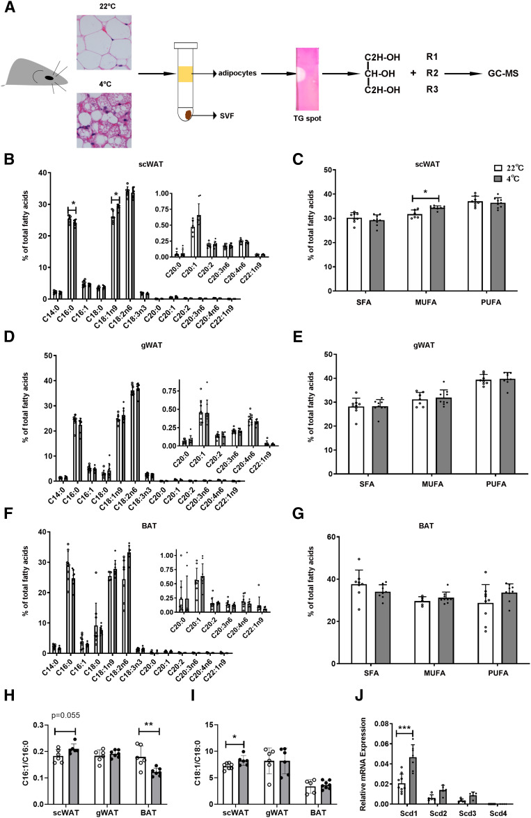
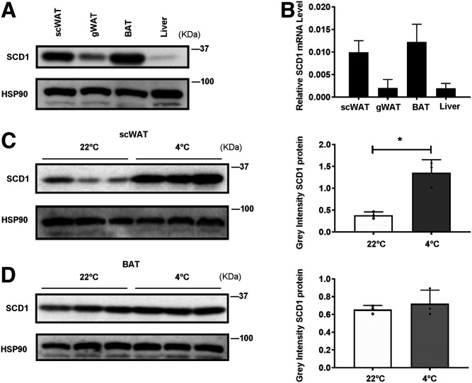
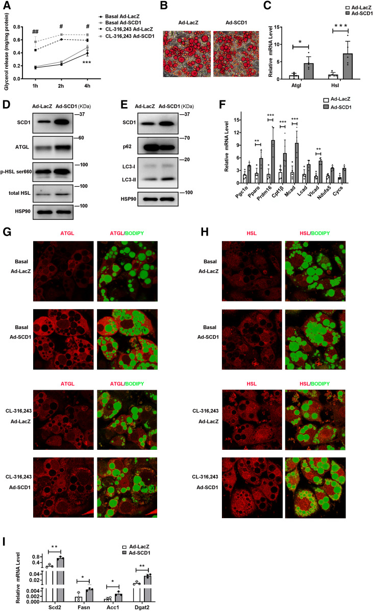
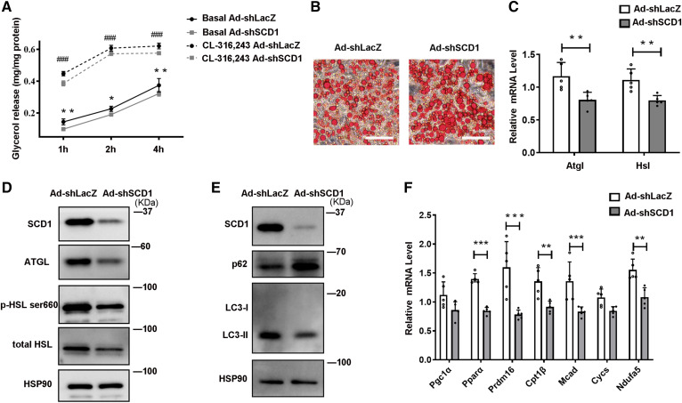
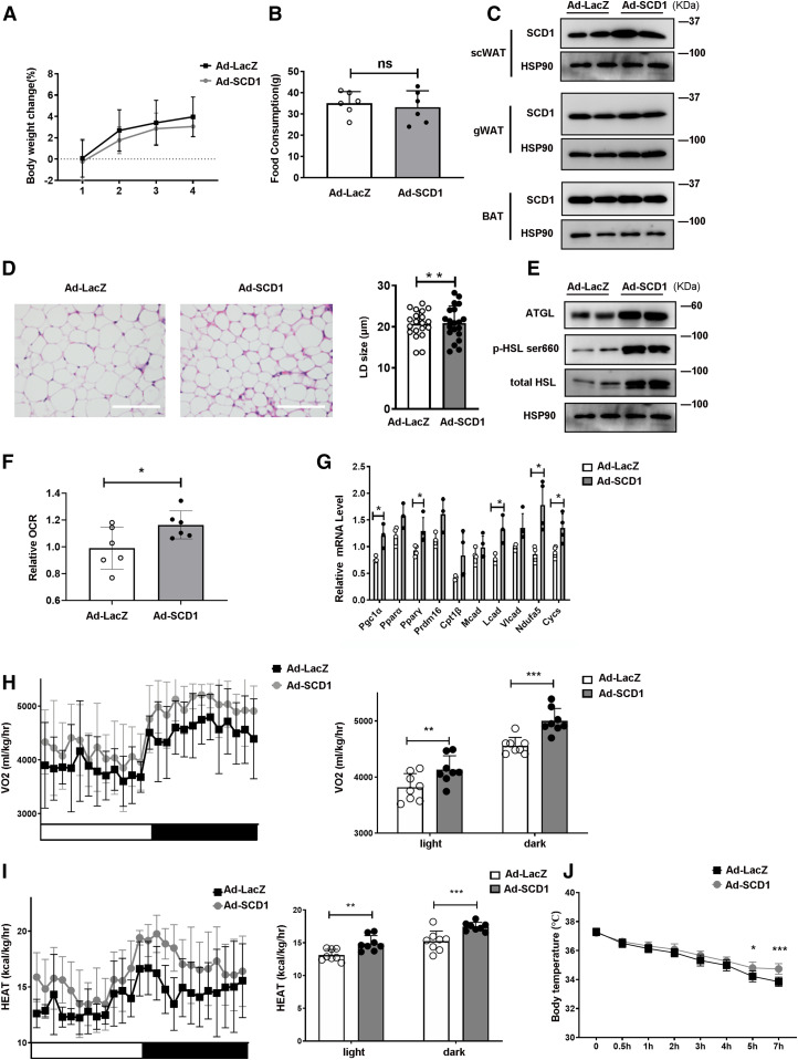
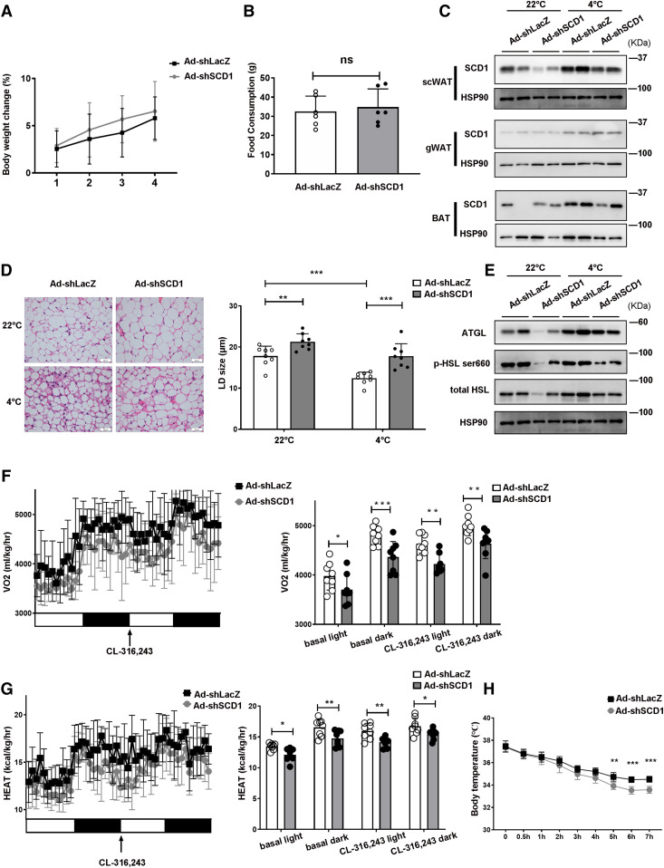
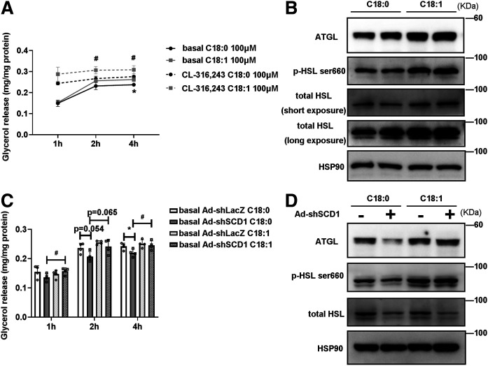
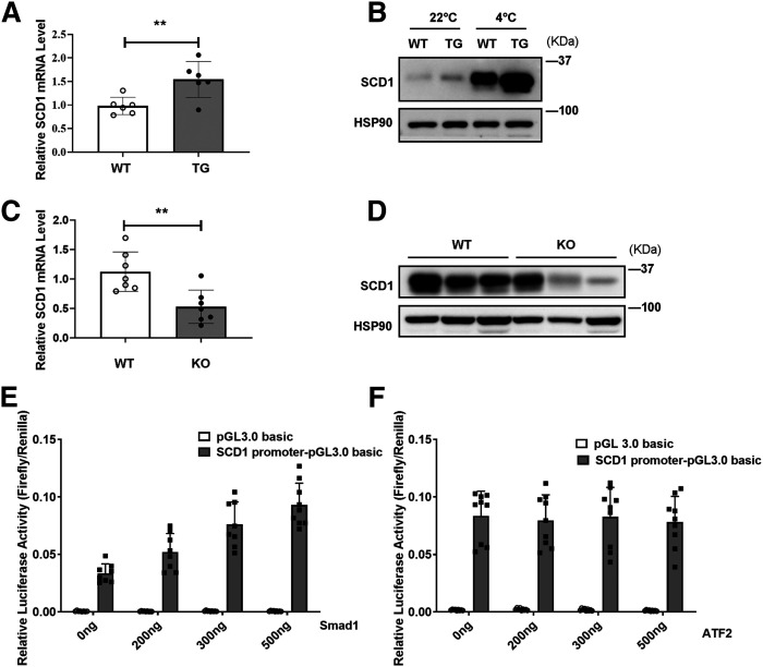
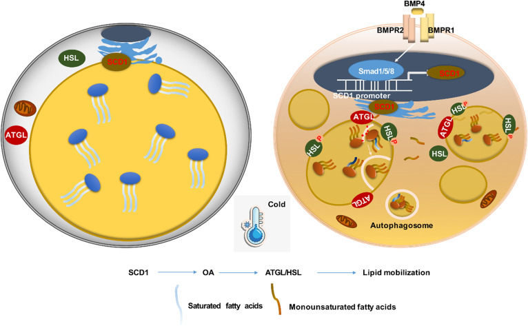
Beiging of white adipose tissue (WAT) has beneficial effects on metabolism. Although it is known that beige adipocytes are active in lipid catabolism and thermogenesis, how they are regulated deserves more explorations. In this study, we demonstrate that stearoyl-CoA desaturase 1 (SCD1) in subcutaneous WAT (scWAT) responded to cold stimulation and was able to promote mobilization of triacylglycerol [TAG (triglyceride)]. In vitro studies showed that SCD1 promoted lipolysis in C3H10T1/2 white adipocytes. The lipolytic effect was contributed by one of SCD1's products, oleic acid (OA). OA upregulated adipose TAG lipase and hormone-sensitive lipase expression. When SCD1 was overexpressed in the scWAT of mice, lipolysis was enhanced, and oxygen consumption and heat generation were increased. These effects were also demonstrated by the SCD1 knockdown experiments in mice. In conclusion, our study suggests that SCD1, known as an enzyme for lipid synthesis, plays a role in upregulating lipid mobilization through its desaturation product, OA.

Keywords: adipocytes; lipolysis; lipophagy; oleic acid; stearoyl-CoA desaturase-1; thermogenesis; triacylglycerol.


Conflict of interest statement

Conflict of interest—The authors declare that they have no conflicts of interest with the contents of this article.

Figures

Fig. 1.
Fig. 1.
The changes of FAs in the TAG of WAT and BAT adipocytes in response to cold exposure. A: Schematic model describes the method of TAG FA in adipocyte analysis. Adipocytes were enzymatically dissociated from scWAT, gWAT, and BAT of C57BL/6J mice either at RT (22°C) or during cold exposure (4°C) for 3 days. TAGs were separated and collected by TLC. After being hydrolyzed by KOH, lipids were esterified to form methyl esters. Methyl esters were detected by GC-MS. Lipid species were quantified based on comprehensive peak area. B, C: FAs were obtained from scWAT of mice housed at RT (22°C) or during cold exposure (4°C) for 3 days. (n = 8 for each group) according the methods in A. FA profiles were analyzed (B) and percentage of SFAs, MUFAs, and PUFAs were calculated (C). D, E: The same analysis as in B and C from gWAT samples of the same mice. F, G: The same analysis as in B and C from BAT samples of the same mice. H, I: SCD desaturation indices were calculated accordingly: SCD-16 = 16:1/16:0 and SCD-18 = 18:1/18:0, as shown in H and I, respectively. J: Relative mRNA expression of Scd1Scd4 in scWAT from mice housed at RT or during cold exposure for 3 days. (n = 7–9 for each group). Statistical analysis: unpaired Student’s t-test in B–J. Data were expressed as mean ± SD. *P < 0.05, **P < 0.01, ***P < 0.001.
Fig. 2.
Fig. 2.
SCD1 is enriched in scWAT and BAT. It is highly induced by cold exposure in adipocytes. A: Tissue distribution of SCD1 protein expression in adipocytes of scWAT, gWAT, BAT, and liver from mice housed at RT (22°C). B: Tissue distribution of Scd1 mRNA expression (relative to 18S rRNA) in adipocytes of scWAT, gWAT, BAT, and liver from mice housed at RT. C: Western blot detection of SCD1 expression in scWAT adipocytes of mice at RT or during cold exposure (4°C) for 3 days (left panel), and the gray density of those bands (right panel) (n = 3 for each group). D: Western blot detection of SCD1 expression in BAT adipocytes of mice at RT or during cold exposure for 3 days (left panel), and the gray density of those bands (right panel) (n = 3 for each group). Statistical analysis: unpaired Student’s t-test in C and D. Data were expressed as mean ± SD. *P < 0.05, **P < 0.01, ***P < 0.001.
Fig. 3.
Fig. 3.
SCD1 promotes lipid mobilization in cultured adipocytes by upregulating levels of lipases, lipophagy, and lipogenesis. C3H10T1/2 mesenchymal stem cells were differentiated for 6 days during the adipogenic program. A: Relative glycerol release from C3H10T1/2 adipocytes treated with Ad-LacZ or Ad-SCD1 for 3 days before the lipolysis experiment. Adipocytes were cultured without or with 0.1 μM CL-316,243 (cell culture medium was changed to phenol red-free medium containing 2% BSA without FAs). The glycerol content of culture medium was quantified with a glycerol release kit (Applygen, E1002) at 1, 2, and 4 h (n = 4 for each group). Total cellular protein was quantified for the glycerol normalization. Experiments were independently repeated three times. B: Oil Red O staining of C3H10T1/2 cells treated with Ad-LacZ or Ad-SCD1. C: Relative mRNA expression of Atgl and Hsl in cells treated with Ad-LacZ or Ad-SCD1 for 3 days (n = 5 in each group). D: Representative Western blot of the lipases (ATGL, total HSL, and HSL phosphorylation) in adipocytes. E: Representative Western blot of the lipophagy markers protein (p62 and LC3-II) in cells treated with Ad-LacZ or Ad-SCD1 for 3 days. F: Relative mRNA expression of the transcriptional regulators and FA oxidation enzymes, nuclear-encoded mitochondrial genes in cells treated with Ad-LacZ or Ad-SCD1 for 3 days (n = 5 in each group). G, H: Immunofluorescence staining with anti-ATGL antibodies (G) and anti-HSL antibodies (H) was performed to reveal localization of endogenous ATGL or HSL in C3H10T1/2 adipocytes treated with Ad-LacZ or Ad-SCD1 for 3 days under basal or 0.1 μM CL-316,243-stimulated 20 min state. LDs were stained with BODIPY 493/503. I: Relative mRNA expression of the lipogenesis genes Scd2, Fasn, Dgat2, and Acc1 in cells treated with Ad-LacZ or Ad-SCD1 for 3 days (n = 3 in each group). Statistical analysis: two-way ANOVA in A, unpaired Student’s t-test in C, F, and I. Data were expressed as mean ± SD. *P indicated for the comparisons at the basal condition (Ad-LacZ vs. Ad-SCD1); #P indicated for the comparisons at the CL-316,243 stimulated condition (Ad-LacZ vs. Ad-SCD1). Data were expressed as mean ± SD. *P < 0.05, **P < 0.01, ***P < 0.001. #P < 0.05, ##P < 0.01, ###P < 0.001.
Fig. 4.
Fig. 4.
Knockdown SCD1 inhibits lipolysis in cultured adipocytes by downregulating lipases and lipophagy. C3H10T1/2 mesenchymal stem cells were differentiated for 6 days during the adipogenic program. A: Relative glycerol release from C3H10T1/2 adipocytes treated with Ad-shLacZ or Ad-shSCD1 for 3 days before the lipolysis experiment. Adipocytes were cultured without or with 0.1 μM CL-316,243 (cell culture medium was changed to phenol red-free medium containing 2% BSA without FAs). The glycerol content of the culture medium was quantified with a glycerol release kit (Applygen, E1002) at 1, 2, and 4 h (n = 4 for each group). Total cellular protein was quantified for the glycerol normalization. Experiments were independently repeated three times. B: Oil Red O staining of C3H10T1/2 cells treated with Ad-shLacZ or Ad-shSCD1. C: Relative mRNA expression of Atgl and Hsl in cells treated with Ad-shLacZ or Ad-shSCD1 for 3 days (n = 5 in each group). D: Representative Western blot of the lipase proteins ATGL, HSL, and p-HSLser660 in cells treated with Ad-shLacZ or Ad-shSCD1 for 3 days. E: Representative Western blot of the lipophagy marker proteins (p62 and LC3-II) in cells treated with Ad-shLacZ or Ad-shSCD1 for 3 days. F: Relative mRNA expression of the transcriptional regulators and FA oxidation enzymes, nuclear-encoded mitochondrial genes in cells treated with Ad-shLacZ or Ad-shSCD1 for 3 days (n = 5 in each group). Statistical analysis: two-way ANOVA in A, unpaired Student’s t-test in C and F. Data were expressed as mean ± SD. *P indicated for the comparisons at the basal condition (Ad-shLacZ vs. Ad-shSCD1); #P indicated for the comparisons at the CL-316,243 stimulated condition (Ad-shLacZ vs. Ad-shSCD1). Data were expressed as mean ± SD. *P < 0.05, **P < 0.01, ***P < 0.001. #P < 0.05, ##P < 0.01, ###P < 0.001.
Fig. 5.
Fig. 5.
Overexpression of SCD1 promotes fat mobilization and thermogenesis in scWAT and enhances whole-body energy expenditure of mice. A: Body weight change of mice during four adenovirus injections (n = 6 for each group). B: Total food consumption of mice injected with adenovirus for 2 weeks (n = 6 in each group). C: Representative Western blots from scWAT, gWAT, and BAT adipocytes of SCD1. D: H&E staining of scWAT from mice injected with Ad-LacZ or Ad-SCD1 adjacently to the scWAT. E: Representative Western blots from scWAT adipocytes of ATGL and HSL. The same samples from the same mouse scWAT in C and E. F: Basal OCR of scWAT was measured and shown (n = 6 in each group). Data were normalized to the Ad-LacZ group. G: Relative mRNA expression of the transcriptional regulators, FA oxidation enzymes, and nuclear-encoded mitochondrial genes in scWAT treated with Ad-LacZ or Ad-SCD1 (n = 4 in each group). H: Whole-body oxygen consumption rate (VO2) of the mice during a 12 h light/12 h dark cycle was measured and the mean of the 12 h light/12 h dark cycle were displayed (n = 8 mice in each group). I: Heat generation of mice in a 12 h light/12 h dark cycle was calculated and the average values for the 12 h light/12 h dark cycle are displayed (n = 8 mice in each group). J: Rectal temperature of mice injected with Ad-LacZ or Ad-SCD1 at 4°C for 7 h (n = 6 for each group). Statistical analysis: unpaired Student’s t-test in A, B, D, F, and G; two-way ANOVA in H, I, and J. Data were expressed as mean ± SD. *P < 0.05, **P < 0.01, ***P < 0.001.
Fig. 6.
Fig. 6.
In mice, SCD1 knockdown inhibits fat mobilization in scWAT lipolysis and decreases whole-body energy expenditure. A: Body weight change of mice during four adenovirus injections (n = 6 for each group). B: Total food consumption of mice injected with adenovirus for 2 weeks (n = 6 in each group). C: Representative Western blots from scWAT, gWAT, and BAT adipocytes of SCD1. D: H&E staining of scWAT from mice injected with Ad-shLacZ or Ad-shSCD1 adjacently to the scWAT. E: Representative Western blots from scWAT adipocytes of ATGL and HSL. The same samples from the same mouse scWAT in C and E. F: Whole-body oxygen consumption rate (VO2) of the mice in basal and CL-316,243 conditions during a 12 h light/12 h dark cycle was measured and the mean in basal and CL-316,243 conditions of the 12 h light/12 h dark cycle were displayed (n = 8 mice in each group). G: Heat generation of mice in basal and CL-316,243 conditions in a 12 h light/12 h dark cycle was calculated and the average values for the 12 h light/12 h dark cycle in basal and CL-316,243 conditions are displayed (n = 8 mice in each group). H: Rectal temperature of mice injected with Ad-shLacZ or Ad-shSCD1 at 4°C for 7 h (n = 6 for each group). Statistical analysis: unpaired Student’s t-test in A, B, and D; two-way ANOVA in F, G. and H. Data were expressed as mean ± SD. *P < 0.05, **P < 0.01, ***P < 0.001.
Fig. 7.
Fig. 7.
OA promotes lipolysis and enhances lipase expression in differentiated C3H10T1/2 adipocytes. A: Relative release of glycerol from C3H10T1/2 adipocytes treated with C18:0 (SA) or C18:1 (OA) at 100 μM for 24 h before the lipolysis experiment. Adipocytes were cultured without or with 0.1 μM of CL-316,243 (cell culture medium was changed to phenol red-free medium containing 2% BSA without FAs). The glycerol content of culture medium was quantified with a glycerol release kit (Applygen, E1002) at 1, 2, and 4 h (n = 4 for each group). Total cellular protein was quantified for the glycerol normalization. Experiments were independently repeated three times. B: Representative Western blots of the lipases (ATGL, total HSL, and p-HSLser660) in cells treated with C18:0 or C18:1 at 100 μM for 24 h. C: Relative release of glycerol from differentiated C3H10T1/2 adipocytes treated with Ad-shLacZ or Ad-shSCD1 for 2 days and then adding 100 μM C18:0 or 100 μM C18:1 for 24 h before the lipolysis experiment. Cell culture medium was changed to phenol red-free medium containing 2% BSA without FA. The glycerol content of culture medium was quantified with a glycerol release kit (Applygen, E1002) at 1, 2, and 4 h (n = 4 in each group). Total cellular protein was quantified for the glycerol normalization. D: Representative Western blots of the lipases (ATGL, total HSL, and p-HSLser660) in cells treated with Ad-shLacZ or Ad-shSCD1 for 2 days and then adding 100 μM C18:0 or 100 μM C18:1 for 24 h. Statistical analysis: two-way ANOVA in A and C. Data were expressed as mean ± SD. In A, *P indicated for the comparisons at the basal condition (100 μM SA vs. 100 μM OA); #P indicated for the comparisons at the CL-316,243-stimulated condition (100 μM SA vs. 100 μM OA). In C, *P indicated for the comparisons at basal C18:0 treatment conditions (Ad-shLacZ vs. Ad-shSCD1); #P indicated for the comparisons at the Ad-shSCD1 treatment conditions (100 μM C18:0 vs. 100 μM C18:1). *P < 0.05, **P < 0.01, ***P < 0.001. #P < 0.05, ##P < 0.01, ###P < 0.001.
Fig. 8.
Fig. 8.
BMP4 participates in promotion of SCD1 expression. A: Relative mRNA expression of Scd1 in scWAT adipocytes of WT and BMP4 TG mice housed at 22°C. B: Representative Western blots of SCD1 protein expression in scWAT adipocytes of WT and BMP4 TG mice housed at RT (22°C) or during cold exposure (4°C) for 3 days. C: Relative mRNA expression of Scd1 in scWAT adipocytes of control group (WT) and Fabp4-Cre-Bmp4LoxP/LoxP (KO) mice housed at RT. D: Representative Western blots of SCD1 protein expression in scWAT adipocytes of WT and BMP4 KO mice housed at RT. E: Luciferase assays for SCD1 promoter constructs and Smad1 transfected into 293T cells. F: Luciferase assays for SCD1 promoter constructs and ATF2 transfected into 293T cells. Statistical analysis: unpaired Student’s t-test in A and C. Data were expressed as mean ± SD. *P < 0.05, **P < 0.01, ***P < 0.001.
Fig. 9.
Fig. 9.
Schematic model highlights the SCD1 function on adipocytes. SCD1 is highly expressed on the endoplasmic reticulum of mature adipocytes isolated from scWAT and catalyzed the desaturation of long-chain SFAs to MUFAs. When exposed to cold or BMP signaling stimulation, SCD1 expression on adipocytes increases and produces more MUFAs, which triggers the browning change in adipocytes. On top of that, one of SCD1 products, OA (C18:1), caused the activation of lipid mobilization, including lipolysis and lipophagy, and further promoted thermogenesis. In conclusion, SCD1 is a new and important regulator of adipocyte lipid mobilization and energy homeostasis.

Similar articles

Cited by

References

    1. Seale P., and Lazar M. A.. 2009. Brown fat in humans: turning up the heat on obesity. Diabetes. 58: 1482–1484. - PMC - PubMed
    1. Fedorenko A., Lishko P. V., and Kirichok Y.. 2012. Mechanism of fatty-acid-dependent UCP1 uncoupling in brown fat mitochondria. Cell. 151: 400–413. - PMC - PubMed
    1. Frontini A., and Cinti S.. 2010. Distribution and development of brown adipocytes in the murine and human adipose organ. Cell Metab. 11: 253–256. - PubMed
    1. Chen K. Y., Cypess A. M., Laughlin M. R., Haft C. R., Hu H. H., Bredella M. A., Enerback S., Kinahan P. E., Lichtenbelt W., Lin F. I., et al. . 2016. Brown Adipose Reporting Criteria in Imaging Studies (BARCIST 1.0): recommendations for standardized FDG-PET/CT experiments in humans. Cell Metab. 24: 210–222. - PMC - PubMed
    1. Xu Z., You W., Zhou Y., Chen W., Wang Y., and Shan T.. 2019. Cold-induced lipid dynamics and transcriptional programs in white adipose tissue. BMC Biol. 17: 74. - PMC - PubMed

LinkOut - more resources