. 2020 Nov 1;205(9):2362-2374.
doi: 10.4049/jimmunol.1901453. Epub 2020 Sep 25.

B Cell Subsets Differentially Contribute to the T Cell-Independent Memory Pool

Affiliations

B Cell Subsets Differentially Contribute to the T Cell-Independent Memory Pool

Christina A Daly et al. J Immunol. .

Abstract

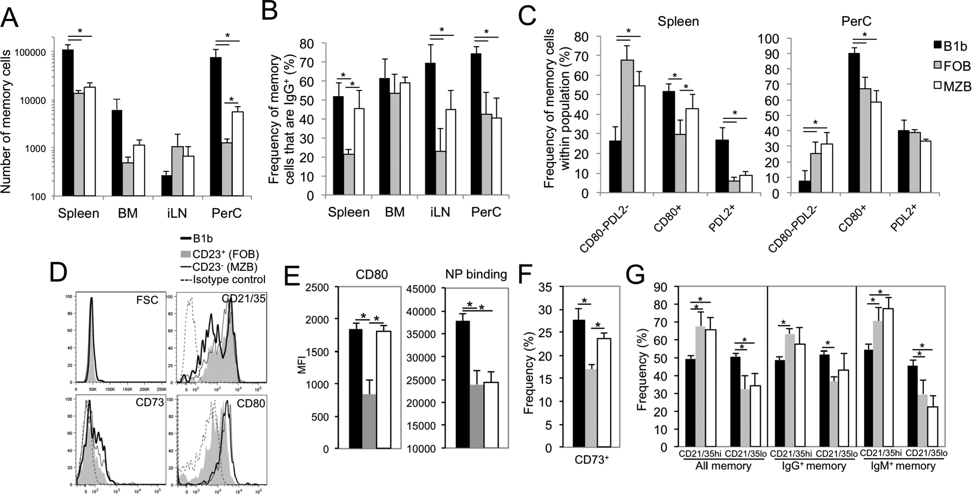
The roles distinct B cell subsets play in clonal expansion, isotype switching, and memory B cell differentiation in response to T cell-independent type 2 Ags (TI-2 Ags) has been understudied. Using sorted B cells from VHB1-8 knock-in mice, we evaluated B-1b, marginal zone, and follicular B cell responses to the TI-2 Ag, NP-Ficoll. All subsets extensively divided in response to NP-Ficoll. Nonetheless, B-1b cells exhibited significantly increased IgG switching and differentiation into Ab-secreting cells (ASC)-a finding that coincided with increased AgR signaling capacity and Blimp1 expression by B-1b cells. All subsets formed memory cells and expressed markers previously identified for T cell-dependent memory B cells, including CD80, PDL2, and CD73, although B-1b cells generated the greatest number of memory cells with higher frequencies of IgG- and CD80-expressing cells. Despite memory formation, secondary immunization 4 wk after primary immunization did not increase NP-specific IgG. However, boosting occurred in B-1b cell-recipient mice when IgG levels declined. CD80+ memory B-1b cells divided, class switched, and differentiated into ASC in response to Ag in vivo, but this was inhibited in the presence of NP-specific IgG. Furthermore, CD80 blockade significantly increased memory B-1b cell division and differentiation to ASC upon Ag restimulation. Collectively, these findings demonstrate B-1b, marginal zone B, and follicular B subsets significantly contribute to the TI-2 Ag-specific memory B cell pool. In particular, we show B-1b cells generate a functional CD80-regulated memory population that can be stimulated to divide and differentiate into ASC upon Ag re-encounter when Ag-specific IgG levels decline.


Figures

Figure 1.
Figure 1.. NP-specific Ab production and memory formation by VHB1–8 Tg B cells
A-C) WT CD45.2+ C57BL/6 recipient mice were given CD43-depleted CFSE-labeled splenic B cells i.v. (4 × 10⁶) and peritoneal B cells i.p. (1.6 × 10⁶) harvested from CD45.1+ VHB1–8 transgenic mice. One day later, mice were immunized with 1 or 25 μg NP40-Ficoll or 25 μg TNP65-Ficoll i.p. A) NP-specific IgMa, IgG, and endogenous IgMb production in recipient mice. B) Frequency and number of VHB1–8 Tg memory B cells (CD45.1+NP-APC+CD19+CD138negCFSElow) in spleen, peritoneal cavity, and bone marrow of recipient mice 21 days post immunization. Right panel indicates frequency of memory B cells which are IgG+ (inclusive of IgG1, IgG2b, and IgG3) among memory cells. C) Representative flow cytometry plots showing CD80 and PDL2 expression by VHB1–8 Tg memory B cells in spleen and peritoneal cavity (left panels) and the frequency of memory B cells that express CD80 (right panel). Asterisks (*) indicate values are significantly different from mice immunized with 25 μg NP-Ficoll (n=4–5 mice/group). Results representative of 2 or more experiments.
Figure 2.
Figure 2.. NP-specific B cell subsets in VHB1–8 Tg mice
A-B) NP-specific B cell frequencies among splenic follicular (FOB), marginal zone (MZB), and peritoneal B-1b cell subsets in VHB1–8 Tg mice. Representative gating strategy is shown in (A) and frequencies of NP12-Fluorescein(Fl)-Ficoll-binding B cells among indicated populations indicated in (B). C-D) Lambda 1 (λ1) light chain expression (C) and NP12-Fl-Ficoll binding levels (D) among VHB1–8 Tg B cell subsets selectively gated for λ1 expression and NP-Ficoll binding, as depicted by the gating within histograms of panels C and A, respectively. Asterisks indicate significant differences between indicated groups (one-way Anova with Tukey’s post-hoc analysis, n=3 mice/group).
Figure 3.
Figure 3.. NP-specific B cell expansion, isotype switching, and differentiation by distinct VHB1–8 Tg B cell subsets 6 days post NP-Ficoll immunization
A-E) B-1b (B220+CD11b+CD5), FOB (CD19+CD21intCD23+CD11b) and MZB (CD19+CD21/35hiCD23lowCD11b) from CD45.1+VHB1–8 Tg mice were FACS-sorted, CFSE labeled and transferred i.v. (5 × 10⁵) into WT recipients. Recipients were immunized with 25 μg NP40-Ficoll on d1. On day 7, tissues were harvested, with CD45.1+ NP-specific CD19+ B cells analyzed by flow cytometry. A) CFSE levels in Tg NP-specific and non-specific splenic B cells recovered from recipient mice. B) CD45.1+ NP-specific B cell frequencies among leukocytes on d7 in recipient spleen, peritoneal cavity (PerC), inguinal lymph node (LN), and bone marrow. C-D) FSC levels for CD45.1+ NP-specific B cells in recipients (C) and CD21/35 expression on donor CD45.1+ NP-specific splenic B cells (D) in immune and naïve recipients. Shaded histograms indicate expression in endogenous (recipient) B cells. E) Frequencies of IgG+, CD138+, and IgG+CD138+ cells among CD45.1+ NP-specific cell populations in spleen, PerC, LN, and bone marrow. Asterisks indicate significant differences between indicated groups (p<0.05, n=3–5/group). Results representative of two or more experiments.
Figure 4.
Figure 4.. Heightened NP-specific Ab production, Ag-induced [Ca2+]i increases, and Blimp1 expression by VHB1–8 Tg B-1b cells
A) B-1b, FOB, and MZB cells from CD45.1+ VHB1–8 Tg mice were FACS-sorted, CFSE labeled and transferred i.v. (5 × 10⁵) into WT recipients. Mice were immunized on d1 and d30 post transfer with 25 μg NP-Ficoll. NP-specific serum IgMa and IgG concentrations were measured by ELISA. Asterisks (*) indicate significant differences compared to FOB group, hash marks (#) indicate significant differences compared to MZB group (p<0.05, n=4–5/group). Results representative of 2 independent experiments. B) NP12-Ficoll-induced [Ca2+]i responses in peritoneal B-1b (CD11b+B220+) and splenic CD23+ and CD23 B220+ B cells from VHB1–8 Tg mice. For baseline readings (first 30 seconds) ([Ca2+]i) values were assessed for Indo-1AM violet/Indo-1AM blue ratios of bulk B cell subsets (ie., both non-Ag- and Ag-specific). Following addition of NP12-Ficoll-fluorescein, NP-binding cells could be visualized and this population (Ag-specific) was gated within each subset to assess changes in ([Ca2+]i) values for NP-Ficoll-fluorescein binding B cells beyond the 30 second time point. Results representative of those obtained from 2–3 mice. C) Blimp1 expression by naïve NP-specific VHB1–8 Tg peritoneal B-1b, splenic FOB and MZB cells. Left panels indicate representative Blimp1 staining (solid line) and control staining with rat IgG PE substituted for Blimp1-PE mAb in intracellular stain for each NP-APC-binding subset. Right graph indicates mean MFI Blimp1 values after subtraction from isotype control values for each individual subset (n=3 mice/group). Results obtained from 2 separate experiments.
Figure 5.
Figure 5.. Generation of memory by distinct VHB1–8 Tg B cell subsets in response to NP-Ficoll
A-C) B-1b, FOB, and MZB from CD45.1+ VHB1–8 Tg mice were FACS-sorted, CFSE-labeled and transferred i.v. (5 × 10⁵) into WT recipients. One day later, mice were immunized with 25 μg NP-Ficoll. A) Number of CD45.1+CD19+VHB1–8 Tg NP-specific memory B cells (CD138negCFSElow) in spleen, inguinal LN, peritoneal cavity, and bone marrow of recipient mice 21 days post immunization. B) Frequency of NP-specific Tg memory B cells in recipient mice that express IgG (inclusive of IgG1, IgG2b, and IgG3). C) Frequency of NP-specific Tg memory B cells in recipient mice that express a CD80PDL2, CD80+ or PDL2+ surface phenotype in spleen and peritoneal cavity. Asterisks (*) indicate significant differences among indicated groups (p<0.05). D-G) Sorted subsets were transferred into WT recipients and mice were immunized with 1 μg NP-Ficoll. On day 40, spleen cells were harvested and memory cells were analyzed for size (FSC) and surface marker expression (D), with NP-APC binding and CD80 MFI (E), frequency of CD73-expressing cells (F), and CD21/35 high and low-expressing population frequencies among all, IgM+, and IgG+ memory B cells (G) quantified. Asterisks (*) indicate significant differences among indicated groups (p<0.05, A-C, n=5–9 recipients/group from 2–3 independent experiments, and D-G, n=3–4 recipients/group).
Figure 6.
Figure 6.. Generation of long-term memory by VHB1–8 Tg B cell subsets in response to NP-Ficoll
B-1b, FOB, and MZB from CD45.1+ VHB1–8 Tg mice were FACS-sorted, CFSE-labeled and transferred i.v. (5 × 10⁵) into WT recipients. One day later, mice were immunized with 25 μg NP-Ficoll. A) Number of CD45.1+CD19+VHB1–8 Tg NP-specific memory B cells (CD138negCFSElow) in spleen and peritoneal cavity of recipient mice 60 days post immunization. Asterisks (*) indicate significant differences among indicated groups (p<0.05, n=4–5/group). Results representative of 2 independent experiments. B) Recipient mice of FACS-sorted peritoneal B-1b and splenic FOB and were immunized with 25 μg NP-Ficoll on d0 and boosted 10 weeks later. NP-specific serum IgM, IgG, and IgMa concentrations were measured by ELISA pre-boost (d70) and post-boost. Asterisks (*) indicate significant differences at the indicated time points compared to preimmune (d70) levels of the same group.
Figure 7.
Figure 7.. Ag-induced expansion and ASC differentiation in CD80+ memory B-1b cells is regulated by CD80 and Ag-specific IgG
A-F) Peritoneal cells enriched for CD19+VHB1–8 Tg memory B-1b cells (CD45.1+ NP-APC+CD11b+CD80) were harvested from donors 2–3 months post NP-Ficoll immunization and either transferred into recipient mice (A-E) or cultured in vitro (F) to assess Ag reactivation. A) Phenotype of CD19+ NP-specific memory B-1b cells (CD45.1+IgM+CD80+CD138negCD11b+) used for transfer experiments. Shaded histograms depict isotype control staining. B-E) CD80+ memory B-1b cells (5 × 10⁵) were transferred into WT (B-D) or mumt (E) mice i.p. as depicted in (A). The next day, mice received 250 μg mouse anti-NP IgG1 or control mouse IgG i.p., and 1 hour later, were immunized with 5 μg NP40-Ficoll i.p. One group received cells without immunization and another received no cells but were immunized. VHB1–8 Tg CD45.1+CD19+NP-APC+ numbers (B) and surface phenotype (C) were assessed by flow cytometry 4 days post immunization. NP-specific serum IgM and IgG (IgG2a, 2c, and 3) concentrations were measured by ELISA for WT (d4; D) and mumt recipients (d6 and 12; E). Asterisks indicate significant differences between immunized mice that received control IgG and naïve or NP-specific IgG1 recipients (p<0.05, n=4 mice/group). F) CFSE-labeled unfractionated peritoneal cells containing VHB1–8 Tg CD45.1+CD19+NP-APC+CD11b+CD80+ memory B-1b cells (as shown in A) or naïve VHB1–8 Tg CD45.1+CD19+NP-APC+ B cells, were cultured in media alone or in the presence of NP-Ficoll and control rat IgG or a CD80 blocking mAb (1G10). On day 4, cell number, division, and phenotype were assessed by flow cytometry. Asterisks indicate significant differences between mean cell numbers (p<0.05). Results are representative of 3 independent experiments.

Similar articles

Cited by

References

    1. Astronomo RD and Burton DR. 2010. Carbohydrate vaccines: developing sweet solutions to sticky situations? Nat. Rev. Drug Discov 9: 308–324. - PMC - PubMed
    1. Wilson D and Braley-Mullen H. 1981. Antigen requirements for priming of type III pneumococcal polysaccharide-specific IgG memory responses: suppression of memory with the T-independent form of antigen. Cell Immunol. 64: 177–186. - PubMed
    1. McMaster PR, Powers KG, Finerty JF and Schiffman G. 1973. Tolerance to type 3 pneumococcal polysaccharide in monkeys. Immunol. Commun 2: 361–370. - PubMed
    1. Poolman J and Borrow R. 2011. Hyporesponsiveness and its clinical implications after vaccination with polysaccharide or glycoconjugate vaccines. Expert Rev. Vaccines 10: 307–322. - PubMed
    1. Brodeur PH and Wortis HH. 1980. Regulation of thymus-independent responses: unresponsiveness to a second challenge of TNP-Ficoll is mediated by hapten-specific antibodies. J. Immunol 125: 1499–1505. - PubMed

Publication types

MeSH terms