. 2020 Sep 25;11(5):e02168-20.
doi: 10.1128/mBio.02168-20.

Rescue of SARS-CoV-2 from a Single Bacterial Artificial Chromosome

Affiliations

Rescue of SARS-CoV-2 from a Single Bacterial Artificial Chromosome

Chengjin Ye et al. mBio. .

Abstract

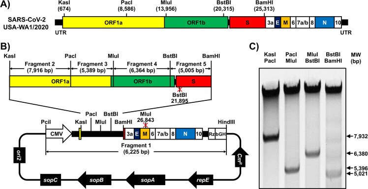
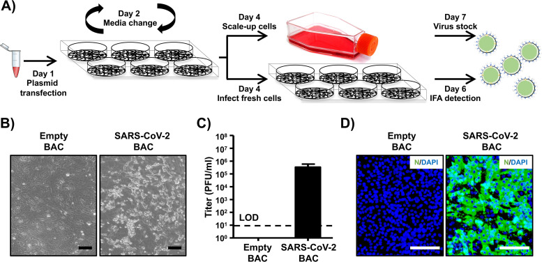
Infectious coronavirus (CoV) disease 2019 (COVID-19) emerged in the city of Wuhan (China) in December 2019, causing a pandemic that has dramatically impacted public health and socioeconomic activities worldwide. A previously unknown coronavirus, severe acute respiratory syndrome CoV-2 (SARS-CoV-2), has been identified as the causative agent of COVID-19. To date, there are no U.S. Food and Drug Administration (FDA)-approved vaccines or therapeutics available for the prevention or treatment of SARS-CoV-2 infection and/or associated COVID-19 disease, which has triggered a large influx of scientific efforts to develop countermeasures to control SARS-CoV-2 spread. To contribute to these efforts, we have developed an infectious cDNA clone of the SARS-CoV-2 USA-WA1/2020 strain based on the use of a bacterial artificial chromosome (BAC). Recombinant SARS-CoV-2 (rSARS-CoV-2) was readily rescued by transfection of the BAC into Vero E6 cells. Importantly, BAC-derived rSARS-CoV-2 exhibited growth properties and plaque sizes in cultured cells comparable to those of the natural SARS-CoV-2 isolate. Likewise, rSARS-CoV-2 showed levels of replication similar to those of the natural isolate in nasal turbinates and lungs of infected golden Syrian hamsters. This is, to our knowledge, the first BAC-based reverse genetics system for the generation of infectious rSARS-CoV-2 that displays features in vivo similar to those of a natural viral isolate. This SARS-CoV-2 BAC-based reverse genetics will facilitate studies addressing several important questions in the biology of SARS-CoV-2, as well as the identification of antivirals and development of vaccines for the treatment of SARS-CoV-2 infection and associated COVID-19 disease.IMPORTANCE The pandemic coronavirus (CoV) disease 2019 (COVID-19) caused by severe acute respiratory syndrome CoV-2 (SARS-CoV-2) is a major threat to global human health. To date, there are no approved prophylactics or therapeutics available for COVID-19. Reverse genetics is a powerful approach to understand factors involved in viral pathogenesis, antiviral screening, and vaccine development. In this study, we describe the feasibility of generating recombinant SARS-CoV-2 (rSARS-CoV-2) by transfection of a single bacterial artificial chromosome (BAC). Importantly, rSARS-CoV-2 possesses the same phenotype as the natural isolate in vitro and in vivo This is the first description of a BAC-based reverse genetics system for SARS-CoV-2 and the first time that an rSARS-CoV-2 isolate has been shown to be phenotypically identical to a natural isolate in a validated animal model of SARS-CoV-2 infection. The BAC-based reverse genetics approach will facilitate the study of SARS-CoV-2 and the development of prophylactics and therapeutics for the treatment of COVID-19.

Keywords: BAC; COVID-19; SARS-CoV-2; coronavirus; hamsters; recombinant virus; reverse genetics.


Figures

FIG 1
FIG 1
Assembly of the SARS-CoV-2 genome into a BAC. (A) Schematic representation of the SARS-CoV-2 genome. The indicated restriction sites were used for cloning the entire viral genome (29,903 nucleotides) of SARS-CoV-2, USA-WA1/2020 strain, into the pBeloBAC11 plasmid. The open reading frames of the viral structural 1a, 1b, spike (S), envelop (E), matrix (M), and nucleocapsid (N) proteins and the accessory (3a, 6, 7a, 7b, 8, and 10) proteins are also indicated. UTR, untranslated regions. Length is not to scale. (B and C) Assembly of the viral genome. (B) The full-length infectious cDNA clone was assembled by sequentially cloning chemically synthesized fragments 1 to 5, which covers the entire viral genome, into the pBeloBAC11 plasmid by using the indicated restriction sites under the control of the cytomegalovirus (CMV) promoter; the clone was flanked at the 3′ end by the hepatitis delta virus (HDV) ribozyme (Rz) and the bovine growth hormone (bGH) termination and polyadenylation sequences. The length of each of the chemically synthesized viral fragments is indicated. Ori2 indicates the origin of the replication of BAC. sopA, sopB, and sopC are the elements to ensure that each bacterial cell gets a copy of the BAC. CmR indicates chloramphenicol resistance. (C) After assembly, the BAC clone harboring the entire viral genome was digested with the indicated restriction enzymes (top), and DNA products were analyzed in a 0.5% agarose gel.
FIG 2
FIG 2
Rescue of rSARS-CoV-2. (A) Schematic representation of the approach followed to rescue rSARS-CoV-2. Vero E6 cells were transiently transfected with the SARS-CoV-2 BAC at day 1. After 24 h, transfection medium was changed to postinfection medium. At day 4, cells were split into T75 flasks and the tissue culture supernatant was used to infect fresh Vero E6 cells. At 48 h postinfection, Vero E6 cells were fixed for detection of rSARS-CoV-2 by immunofluorescence, and the tissue culture supernatant of the scaled-up Vero E6 cells was collected at 72 h. As an internal control for this experiment, Vero E6 cells were transfected with the empty BAC. (B) CPE. Images of empty or SARS-CoV-2 BAC-transfected Vero E6 cells at 72 h posttransfection are shown. Scale bars, 100 μm. (C) Viral titers. Tissue culture supernatant from mock-infected (empty BAC) or transfected Vero E6 cells in T75 flasks was collected and titrated by immunofluorescence. Data are presented as means ± SDs. LOD, limit of detection. (D) IFA. Vero E6 cells infected with the tissue culture supernatants from transfected Vero E6 cells were fixed at 48 h postinfection, and viral detection was carried out by using a SARS-CoV cross-reactive monoclonal antibody (1C7) against the N protein (green). Cellular nuclei were stained by 4′,6-diamidino-2-phenylindole (DAPI; blue). Scale bars, 100 μm.
FIG 3
FIG 3
Characterization of rSARS2-CoV-2 in vitro. (A and B) Genotypic characterization. Vero E6 cells were mock infected or infected (MOI, 0.01) with rSARS-CoV-2 or the SARS-CoV-2 USA-WA1/2020 natural isolate. At 24 h postinfection, total RNA from Vero E6 cells was extracted and a 1,297-bp region of the M gene (nt 26488 to 27784) was amplified by RT-PCR. Amplified DNA was subjected to MluI digestion (Fig. 1). (A) Undigested (top) and digested (bottom) samples were separated in a 0.7% agarose gel. The RT-PCR-amplified DNA product was also sequenced to verify the presence of the silent mutation in the MluI restriction site introduced in the viral genome of the rSARS-CoV-2 (Fig. 1). (B) The MluI restriction site is underlined in red, and the silent mutation introduced to remove the MluI restriction site (T to A) is shown in the black box. (C) Verification of BAC and rSARS-CoV-2 sequences. The SARS-CoV-2 non-reference allele frequency was calculated by comparing short reads to the reference genome of the USA-WA1/2020 reference. All variants were at low frequency in the P6 natural isolate (top), the BAC (bottom), and rSARS-CoV-2 (middle), with the exception of introduced variants at positions 21895 and 26843, which were fixed in the BAC and in rSARS-CoV-2. Non-reference alleles present in less than 1% of reads are not shown. (D) Plaque phenotype. Vero E6 cells were infected with ∼20 PFU of rSARS-CoV-2 (left) or the natural SARS-CoV-2 isolate (right). After 72 h of incubation at 37°C, cells were fixed and immunostained with the N protein 1C7 monoclonal antibody. (E) Growth kinetics. Vero E6 cells were infected (MOI, 0.01) with rSARS-CoV-2 or the natural SARS-CoV-2 isolate. At the indicated times postinfection, tissue culture supernatants were collected and viral titers were assessed by plaque assay (PFU/ml). Data are presented as means ± SDs. LOD, limit of detection.
FIG 4
FIG 4
Pathogenicity of rescued rSARS-CoV-2 in vivo. Golden Syrian hamsters were mock infected (n = 2) or infected (n = 6) with 2 × 104 PFU of rSARS-CoV-2 or SARS-CoV-2. (A) Gross pathological lung lesions. Animals were euthanized at 2 and 4 days postinfection, and lungs from mock-infected (i and iv) or infected (rSARS-CoV-2 [ii and v] and SARS-CoV-2 [iii and vi]) animals were observed for gross pathological changes, including congestion and atelectasis (white arrows) and frothy trachea exudates (black arrows). Scale bars, 1 cm. (B) Macroscopic pathology scoring analysis. Distributions of pathological lesions, including consolidation, congestion, and atelectasis, were measured using ImageJ and are represented as percentages of the total lung surface area. ns, not significant. (C and D) Virus titers. Viral titers in the lungs (C) and nasal turbinates (D) of rSARS-CoV-2- and SARS-CoV-2-infected golden Syrian hamsters were determined at days 2 and 4 postinfection (3 hamsters per time point). Data are means ± SDs. ns, not significant.

Update of

References

    1. Lopez-Ortiz E, Lopez-Ortiz G, Mendiola-Pastrana IR, Mazon-Ramirez JJ, Diaz-Quinonez JA. 2020. From the handling of an outbreak by an unknown pathogen in Wuhan to the preparedness and response in the face of the emergence of Covid-19 in Mexico. Gac Med Mex 156:132–137. doi:10.24875/GMM.M20000346. - DOI - PubMed
    1. Ralph R, Lew J, Zeng TS, Francis M, Xue B, Roux M, Ostadgavahi AT, Rubino S, Dawe NJ, Al-Ahdal MN, Kelvin DJ, Richardson CD, Kindrachuk J, Falzarano D, Kelvin AA. 2020. 2019-nCoV (Wuhan virus), a novel Coronavirus: human-to-human transmission, travel-related cases, and vaccine readiness. J Infect Dev Ctries 14:3–17. doi:10.3855/jidc.12425. - DOI - PubMed
    1. Khan S, Nabi G, Han G, Siddique R, Lian S, Shi H, Bashir N, Ali A, Shereen MA. 2020. Novel coronavirus: how things are in Wuhan. Clin Microbiol Infect 26:399–400. doi:10.1016/j.cmi.2020.02.005. - DOI - PMC - PubMed
    1. Stoye E. 7 April 2020. The pandemic in pictures: how coronavirus is changing the world. Nature doi:10.1038/d41586-020-01048-7. - DOI - PubMed
    1. Qian X, Ren R, Wang YF, Guo Y, Fang J, Wu Z-D, Liu P-L, Han T-R, Members of Steering Committee, Society of Global Health, Chinese Preventive Medicine Association. 2020. Fighting against the common enemy of COVID-19: a practice of building a community with a shared future for mankind. Infect Dis Poverty 9:34. doi:10.1186/s40249-020-00650-1. - DOI - PMC - PubMed