. 2020 Sep 25;10(1):15765.
doi: 10.1038/s41598-020-71550-5.

Efficiency of the four proteasome subtypes to degrade ubiquitinated or oxidized proteins

Affiliations

Efficiency of the four proteasome subtypes to degrade ubiquitinated or oxidized proteins

Joanna Abi Habib et al. Sci Rep. .

Abstract

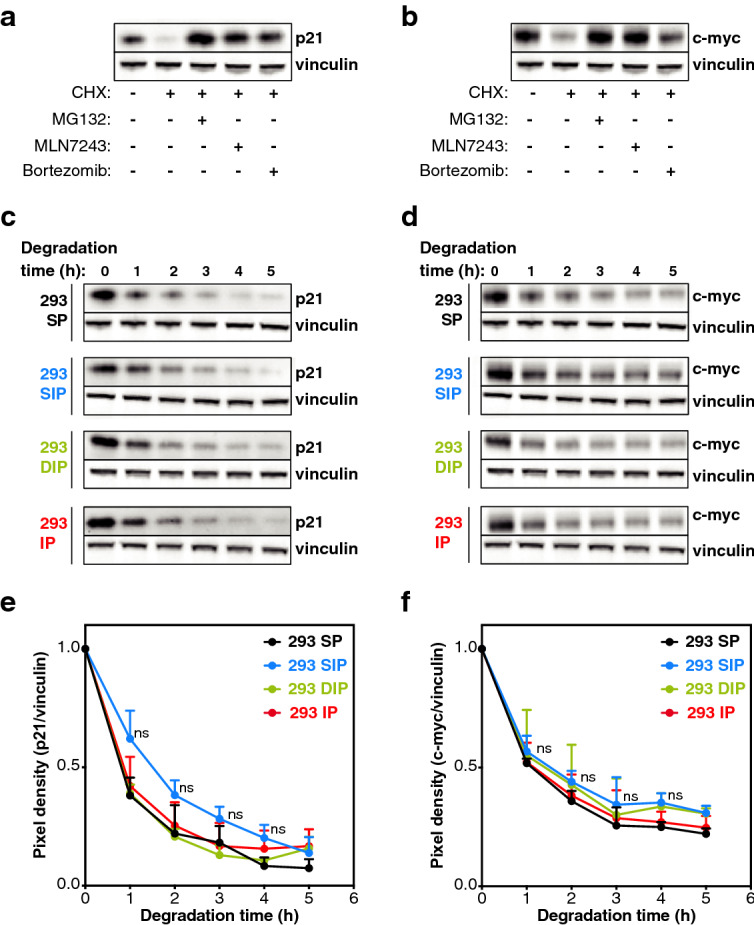
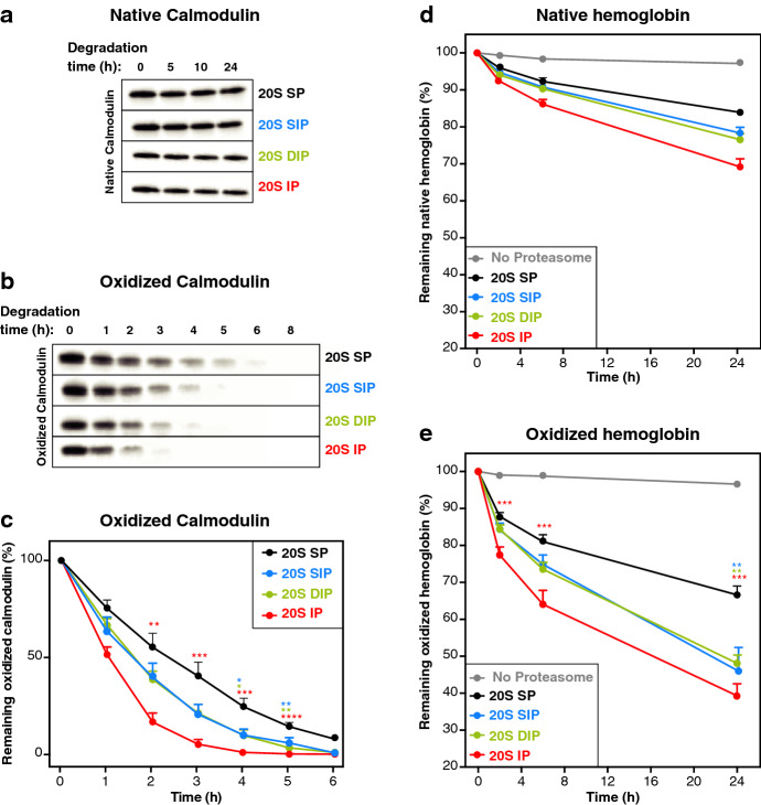
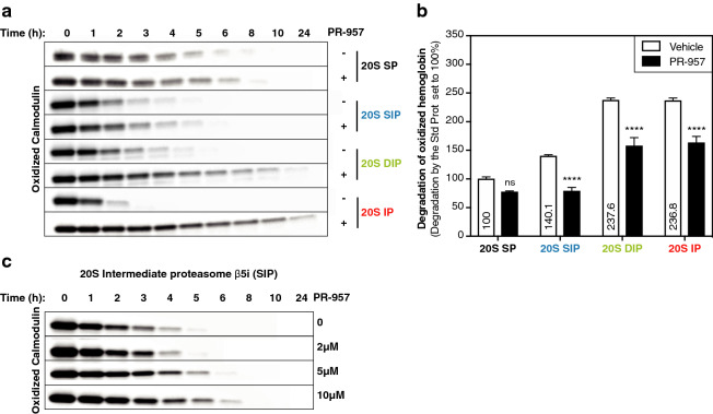
The proteasome is responsible for selective degradation of proteins. It exists in mammalian cells under four main subtypes, which differ by the combination of their catalytic subunits: the standard proteasome (β1-β2-β5), the immunoproteasome (β1i-β2i-β5i) and the two intermediate proteasomes (β1-β2-β5i and β1i-β2-β5i). The efficiency of the four proteasome subtypes to degrade ubiquitinated or oxidized proteins remains unclear. Using cells expressing exclusively one proteasome subtype, we observed that ubiquitinated p21 and c--myc were degraded at similar rates, indicating that the four 26S proteasomes degrade ubiquitinated proteins equally well. Under oxidative stress, we observed a partial dissociation of 26S into 20S proteasomes, which can degrade non-ubiquitinated oxidized proteins. Oxidized calmodulin and hemoglobin were best degraded in vitro by the three β5i-containing 20S proteasomes, while their native forms were not degraded. Circular dichroism analyses indicated that ubiquitin-independent recognition of oxidized proteins by 20S proteasomes was triggered by the disruption of their structure. Accordingly, β5i-containing 20S proteasomes degraded unoxidized naturally disordered protein tau, while 26S proteasomes did not. Our results suggest that the three β5i-containing 20S proteasomes, namely the immunoproteasome and the two intermediate proteasomes, might help cells to eliminate proteins containing disordered domains, including those induced by oxidative stress.


Conflict of interest statement

The authors declare no competing interests.

Figures

Figure 1
Figure 1
Degradation of ubiquitinated proteins by the standard proteasome (SP), the intermediate proteasome β5i (SIP), the intermediate proteasome β1i–β5i (DIP) and the immunoproteasome (IP). (a) p21 and (b) c-myc are degraded in a proteasome and ubiquitin-dependent manner. Western blot analysis of lysates of 293 SP cells treated or not with cycloheximide (CHX) alone or in combination with MLN7243, an inhibitor of the ubiquitin-activating enzyme, or with MG132 or bortezomib, two proteasome inhibitors. Cells were treated with MLN7243, MG132 or Bortezomib 30 min prior to the 5 h treatment with cycloheximide. (c, d) Western blot analysis of the kinetics of degradation of (c) p21 and (d) c-myc in 293 cell lines expressing the different proteasome subtypes. (e, f) Densitometric evaluation of the kinetics of the degradation of (e) p21 and (f) c-myc in the four different cell lines. All values (+ SD) are collected from three independent experiments. Full-length images for (a-d) are presented in Fig. S10.
Figure 2
Figure 2
Oxidative stress dissociates 26S proteasomes into free 20S proteasomes. (a) Relative abundance of the different proteasome regulators and ECM29 bound to 20S proteasomes under oxidative stress induced by H2O2. 293 SP cells were treated with or without 2 mM of H2O2 for 30 min, proteins were cross-linked in vivo with formaldehyde, proteasomes were immuno-purified using the anti-α2 antibody (MCP21) and the different proteasome regulators were analyzed using label-free quantitative MS along with ECM29. The abundance of 20S interactors were measured in control and oxidized conditions, normalized to the abundance of 20S, and then the oxidized condition was normalized to the control condition. All values are means of three independent experiments + SD. (b) Pie chart showing the proportion of free 20S proteasome and of the different regulators associated to 20S proteasomes under control conditions (left panel) and oxidative stress (right panel).
Figure 3
Figure 3
More efficient degradation of oxidized proteins by the 20S immunoproteasome (IP) and the two 20S intermediate proteasomes β5i (SIP) and β1i­-β5i (DIP). (ac) Western blot analysis of the kinetics of degradation of (a) native and (b) oxidized calmodulin by the four 20S proteasome subtypes. (c) Densitometric evaluation of the kinetics of the degradation of the oxidized calmodulin by the four proteasome subtypes. All values (+ SEM) are collected from seven independent experiments using three different batches of purified proteasomes. (d, e) Kinetics of degradation of tritium-labeled (d) native and (e) oxidized hemoglobin. These two forms of hemoglobin were incubated with the four 20S proteasome subtypes. To monitor protein degradation, samples were collected at different time points, precipitated with trichloroacetic acid (TCA) and the radioactivity present in the supernatant was measured. Remaining hemoglobin was quantified by subtracting the radioactivity measured in the supernatant from the total radioactivity. Proteins were oxidized by 24 h incubation in 50 mM H2O2. All values are means (+ SEM) of seven independent experiments using three different batches of purified proteasomes. Full-length images for (a-b) are presented in Fig. S11.
Figure 4
Figure 4
Degradation of oxidized proteins is triggered by disruption of their structure. (a) Analysis by circular dichroism of the secondary structure of native , calmodulin, oxidized calmodulin and oxidized calmodulin treated with 0.5 mM CaCl2. The top panel shows the circular dichroism spectra and the bottom panel shows percentages of the different secondary structures obtained by analyzing the circular dichroism spectra using the Contin-LL program. (b) The left panel illustrates a western blot showing that CaCl2 treatment of oxidized calmodulin reduced its degradation by 20S immunoproteasomes. These results are representative of three independent experiments. The right panel shows that CaCl2 did not affect the activity of 20S immunoproteasomes, as measured with fluorogenic substrate Suc-LLVY-AMC. (c) Analysis by circular dichroism of the secondary structure of native hemoglobin and oxidized hemoglobin. The top panel shows the circular dichroism spectra and the bottom panel shows percentages of the different secondary structures. (d) Kinetics of degradation of labeled unoxidized and oxidized hemoglobin. These two forms of hemoglobin were incubated with the 20S IP, and protein degradation was monitored as described in Fig. 3e. Similar results were obtained from seven independent experiments. (e, f) Assessing the level of surface exposed hydrophobic patches in the three forms of calmodulin (e) and in the two forms of hemoglobin (f) using the Nile Red probe. Nile Red was incubated with the different samples and its fluorescence at 630 nm, which increases in a non-polar environment, was measured. Relative fluorescence units (RFU) at 630 nm of each sample was reported to the RFU at 630 nm of the corresponding native protein, and the absolute RFU values at 630 nm of the native proteins are indicated on the histogram. All values are mean + SEM of three independent experiments. Full-length images for (b) are presented in Fig. S12.
Figure 5
Figure 5
The presence of oxidized residues does not explain the differential degradation of oxidized proteins by the four 20S proteasome subtypes. Assay comparing the fluorescence emitted following the degradation of oxidized and unoxidized FRET (Fluorescence resonance energy transfer)-peptides by the four proteasome subtypes. Oxidized and unoxidized precursor peptides were derived from (a) calmodulin, (b) hemoglobin, (c) NY-ESO1 and (d) LCMV nucleoprotein. Unoxidized precursor peptides contained a methionine residue which was replaced by methionine sulfoxide (Mox) in the corresponding oxidized precursor. These precursor peptides were incubated with the four proteasome subtypes, and their degradation was monitored by measuring fluorescence at 420 nm. RFU at 420 nm of each sample was reported to the RFU at 420 nm of the unoxidized precursor degraded by the SP. The absolute RFU values at 420 nm of the unoxidized precursors degraded by the four proteasome subtypes are indicated on the histogram. All values are mean + SEM of three independent experiments.
Figure 6
Figure 6
Catalytic immuno-subunit β5i plays an important role in the degradation of oxidized proteins by β5i-containing 20S proteasomes. (a) Western blot analysis of the kinetics of degradation of oxidized calmodulin by the four proteasome subtypes in the presence or absence of the β5i-specific inhibitor PR-957 (1 μM). These results are representative of three independent experiments. (b) Assessing the degradation of the radiolabeled oxidized hemoglobin by the four proteasome subtypes in the presence or in the absence of the β5i-specific inhibitor PR-957 after 24 h of digestion. The percentage of degraded hemoglobin was reported in each case to the percentage of degraded hemoglobin by the SP in the absence of PR-957, and the absolute percentages of degraded hemoglobin in the vehicle condition are indicated on the histogram. Error bars are SEM of three independent experiments. (c) Western blot analysis of the kinetics of degradation of oxidized calmodulin by the intermediate proteasome β5i treated with increased concentrations of the PR-957 inhibitor. These results are representative of three independent experiments. Full-length images for (a) and (c) are presented in Fig. S13.
Figure 7
Figure 7
More efficient degradation of intrinsically disordered protein tau by 20S proteasomes containing subunit β5i. (a) Western blot analysis of the kinetics of degradation of recombinant tau 2N4R by the four 20S proteasome subtypes. (b) Densitometric evaluation of the kinetics of the degradation of tau by the four 20S proteasome subtypes. All values (+ SEM) are collected from three independent experiments. (c) Western blot analysis of the kinetics of degradation of tau by the four 26S proteasome subtypes. These results are representative of three independent experiments. Full-length images for (a) and (c) are presented in Fig. S14.

References

    1. Dikic I. Proteasomal and autophagic degradation systems. Annu. Rev. Biochem. 2017;86:193–224. doi: 10.1146/annurev-biochem-061516-044908. - DOI - PubMed
    1. Dengjel J, et al. Autophagy promotes MHC class II presentation of peptides from intracellular source proteins. Proc. Natl. Acad. Sci. USA. 2005;102:7922–7927. doi: 10.1073/pnas.0501190102. - DOI - PMC - PubMed
    1. Rock KL, et al. Inhibitors of the proteasome block the degradation of most cell proteins and the generation of peptides presented on MHC class I molecules. Cell. 1994;78:761–771. doi: 10.1016/S0092-8674(94)90462-6. - DOI - PubMed
    1. Groll M, et al. Structure of 20S proteasome from yeast at 2.4 A resolution. Nature. 1997;386:463–471. doi: 10.1038/386463a0. - DOI - PubMed
    1. Hisamatsu H, et al. Newly identified pair of proteasomal subunits regulated reciprocally by interferon gamma. J. Exp. Med. 1996;183:1807–1816. doi: 10.1084/jem.183.4.1807. - DOI - PMC - PubMed

Publication types

MeSH terms

Substances