. 2020 Sep 25;10(1):15808.
doi: 10.1038/s41598-020-72875-x.

Myh11+ microvascular mural cells and derived mesenchymal stem cells promote retinal fibrosis

Affiliations

Myh11+ microvascular mural cells and derived mesenchymal stem cells promote retinal fibrosis

H Clifton Ray et al. Sci Rep. .

Abstract

Retinal diseases are frequently characterized by the accumulation of excessive scar tissue found throughout the neural retina. However, the pathophysiology of retinal fibrosis remains poorly understood, and the cell types that contribute to the fibrotic response are incompletely defined. Here, we show that myofibroblast differentiation of mural cells contributes directly to retinal fibrosis. Using lineage tracing technology, we demonstrate that after chemical ocular injury, Myh11+ mural cells detach from the retinal microvasculature and differentiate into myofibroblasts to form an epiretinal membrane. Inhibition of TGFβR attenuates Myh11+ retinal mural cell myofibroblast differentiation, and diminishes the subsequent formation of scar tissue on the surface of the retina. We demonstrate retinal fibrosis within a murine model of oxygen-induced retinopathy resulting from the intravitreal injection of adipose Myh11-derived mesenchymal stem cells, with ensuing myofibroblast differentiation. In this model, inhibiting TGFβR signaling does not significantly alter myofibroblast differentiation and collagen secretion within the retina. This work shows the complexity of retinal fibrosis, where scar formation is regulated both by TGFβR and non-TGFβR dependent processes involving mural cells and derived mesenchymal stem cells. It also offers a cautionary note on the potential deleterious, pro-fibrotic effects of exogenous MSCs once intravitreally injected into clinical patients.


Conflict of interest statement

P.A.Y. is the founder of RetiVue, LLC, a company that develops innovative, affordable, and convenient retinal screening technologies for patients. P.A.Y. is also a consultant of Genentech/Roche. All other authors declare no competing interests.

Figures
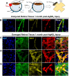
Figure 1
Figure 1
Endogenous Myh11+ mural cells on the retinal microvasculature exhibit a myofibroblast phenotype after a chemical burn to the murine sclera. (A) Model demonstrating that silver-nitrate-burn injury to the sclera induced formation of retinal fibrotic scar tissue. (B) Immunostained, uninjured retinal tissue revealed Myh11+ mural cells, labeled by tdTomato, are found only on the CD31+ retinal microvasculature. Col-IV is expressed only in the basement membrane of the retinal microvasculature. Scale bar, 15 µm. (C) Immunostained retinas one-month post-burn injury showed multiple off-vessel Myh11+ mural cells (tdTomato+), which is indicated by the lack of overlap with the blood vessel endothelium marker CD31. (D,E) Off-vessel Myh11+ mural cells display αSMA+ stress fibers and are positive for Col-IV, Col-III, and F-actin as shown by fluorescently labeled phalloidin. Scale bar, 15 µm. Animals were tested beginning at 10–12 weeks of age. Immunohistochemistry images are representative of three uninjured and injured eyes of Myh11-tdTomato mice. Field of view in injured eyes were selected based on the visual observation of off-vessel Myh11+ mural cells.
Figure 2
Figure 2
Intravitreal injection of the TGFβR inhibitor, SB431542, reduces retinal scar formation in Myh11-tdTomato mice 1-month post chemical burn injury. (A) A Luminex multiplex assay demonstrated TGFβ1 and CXCL10 concentrations were significantly increased in injured neural retina of Myh11-tdTomato mice 1-month post chemical injury (n = 4 paired eyes). The assay also demonstrated IL-1a, IL-2, IL-4, and IL-17 concentrations were significantly decreased in the neural retina of 1-month-post chemically burned eyes as compared to the contralateral uninjured eyes (n = 4 paired eyes). Data are represented as mean ± standard error of mean (SEM). (B) Experimental design for global inhibition of the TGFβ pathway in the eye after chemical-burn injury, where injured eyes received intravitreal injections of either 100 µm SB431542 or carrier control at both 7 and 21 days post injury. (C) Representative images display captured tile scans of immunostained retinas harvested 30 days post-injury from eyes intravitreally injected with vehicle control or 100 µm SB431542. Retinal scar formation was revealed by fibrotic Col-IV and αSMA+ stress fibers. Scale bar, 1000 µm. (D) Scar area generated by the chemical burn [white outline in (C)] was quantified via ImageJ tracing tool, and analysis showed SB431542 significantly decreased retinal fibrosis as compared to contralateral vehicle control injected eyes (n = 10 paired eyes). Animals were tested beginning at 10–12 weeks of age. *p < 0.05, **p < 0.01. Data were analyzed using a ratio paired t test (A), or Wilcoxon test (D).
Figure 3
Figure 3
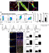
Adipose-derived, lineage-marked Myh11+ mural cells give rise to mesenchymal stem cells (MSCs) during adaptation and growth in vitro. (A) Immunostained epididymal adipose tissue from Myh11-eYFP mice indicated eYFP+ (green) lineage marker is expressed in microvascular smooth muscle cells (vSMCs) (arrowhead) and microvascular pericytes (PCs) (asterisk) along lectin + blood vessels. Scale bar, 50 µm. (B) Immunostained adipose tissue revealed vSMC “tire tread” pattern on larger arterioles. Scale bar, 25 µm. (C) PCs are wrapped around adipose capillary microvasculature. Scale bar, 10 µm. (D) Flow cytometry analysis showed adipose Myh11+ mural cells collected from the SVF have relatively low endogenous expression of CD73, CD90, CD105, and CD146, however, after isolation via fluorescence activated cell-sorting (FACS), cultured, passage 3–5 Myh11+ mural cells significantly increased expression of CD73, CD90, CD105, and CD146 in vitro (three independent flow analyses per panel). (E) Graphical representation of flow cytometry analysis demonstrated significant increase of MSC surface antigens in Myh11+ mural cells after isolation from the SVF and cultured in vitro. (F) Flow cytometry analysis also revealed FAC-sorted and cultured passage 3–5 Myh11+ mural cells lacked expression for hematopoetic, endothelial, and macrophage markers CD11b, CD19, CD34, CD31, and CD45 (three independent flow analyses per panel). (G,H) Protein and genetic analysis of passage 2 Myh11+ mural cells when cultured in adipogeneic, chondrogenic, or osteogenic media for 14 days. (G) Increase in FABP4, Collagen II, and Osteopontin was observed by immunohistochemistry in Myh11+ mural cells undergoing tri-differentiation. Scale bar, 50 µm. (H) qPCR showed mRNA expression of protein markers and transcription factors involved in adipogenesis, chondrogenesis, and osteogenesis were significantly upregulated in Myh11+ mural cells following tri-differentiation (n = 3 biological replicates). Relative expression is normalized to GAPDH expression in each sample. Results are represented as mean ± standard error of mean (SEM). Data were analyzed using multiple unpaired t tests followed by the Holm–Sidak post-hoc comparisons to correct for multiple comparisons (E), or a ratio paired t-test (H). *p < 0.05, **p < 0.01, ***p<0.001. Immunohistochemistry images were captured through randomly sampling of microvasculature tissue and culture wells. Tissue and cultured cells were isolated from Myh11-eYFP mice 10–12 weeks of age.
Figure 4
Figure 4
Intravitreally injected Myh11-derived MSCs accelerate microvasculature recovery and adopt a perivascular position during murine oxygen-induced retinopathy (OIR). (A) Diagram illustrating the timeline of the murine OIR model and intravitreal injection of Myh11-derived MSCs. After hyperoxia injury from P7 to P12, pup eyes were intravitreally injected with PBS-vehicle or 10,000 Myh11-derived MSCs at passage 3–5, and analyzed at P14 and P17 post-injection. (B) At P14 and P17, intravitreally injected Myh11-derived MSCs (DiI+/eYFP+) were able to integrate into the retinal tissue and associate with Col-IV+ retinal vasculature. Scale bar, 10 µm. (C) Representative immunostained retinal flatmounts at P14 and P17 are shown with outlined area (yellow) representing capillary dropout region caused by OIR. Retinal blood vessels were immunostained with lectin (red). Quantification of capillary dropout in ImageJ showed eyes intravitreally injected with Myh11-derived MSCs experienced a significant reduction in capillary dropout area at P14 (n = 7 paired eyes), and at P17 (n = 10 paired eyes) when compared to the contralateral PBS vehicle control eyes. Scale bar, 1000 µm. *(p < 0.05). All data were analyzed using a Wilcoxon test.
Figure 5
Figure 5
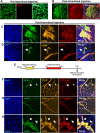
Within the murine OIR model, intravitreal injected Myh11-derived MSCs in the vitreous gel exhibit a myofibroblast phenotype, while endogenous, retinal Myh11+ mural cells remain in a perivascular position. (A) Immunostained Myh11-derived MSCs lacked expression of Col-IV in vitro, however, (B) immunostained retinas revealed intravitreal injected Myh11-derived MSCs expressed Col-IV in the vitreous gel, which formed a dense, fibrotic pre-retinal membrane in murine OIR eyes. Scale bars, 200 µm. (C) Intravitreal injected passage 3–5 Myh11-derived MSCs expressed αSMA+ stress fibers and Col-IV, and (D) Myh11-derived MSCs have reduced expression of Myh11 following injection (arrow) compared to the endogenous Myh11 expressed in retinal mural cells. DAPI stained nuclei of underlying retinal ganglion cells in addition to injected MSCs. Scale bars, 100 µm. (E) Experimental design where tamoxifen is delivered postnatal day 1–3 Myh11-tdTomato mice to induce expression of tdTomato in Myh11+ mural cells. Induced mice are then exposed to hyperoxia from postnatal day 7–12 to cause OIR injury, with retinas harvested at P17 to determine cell fate of endogenous, retinal Myh11+ mural cells. (F) At P17, endogenous, retinal Myh11+ mural cells resided on Col-IV+/CD31+ vessels, with αSMA expression higher in vSMCs (arrow) than PCs (asterisk). Scale bar, 100 µm. (G) Myh11+ mural cells remained on vessel with no vSMCs-PCs found off vessel. Scale bar, 100 µm. (H) Neither retinal vSMCs or PCs extended processes from CD31 tip cells (arrow) at the leading front of the regenerating retinal microvasculature. Scale bar, 25 µm. Immunohistochemistry images represent fields of view that were sampled based on the presence of eYFP and tdTomato expression within culture and tissue. Images are also representative of at least three biological replicates or animals.
Figure 6
Figure 6
Smad4 knockdown within Myh11-derived MSCs does not abolish induction of proliferative vitreoretinopathy following intravitreal injection of these cells within the murine OIR model. (A) A Luminex multiplex assay demonstrated no significant difference in active TGFβ1 protein concentration within the neural retina and vitreous of P14 wildtype (WT) mice and WT mice that underwent OIR (n = 5 unpaired eyes). Cropped Western blot from representative lanes of one gel (B) and densitometry quantification (C) demonstrated significant knockdown of Smad4 in Myh11-derived MSCs through mSmad4-shRNA adenovirus vectors (n = 6 biological replicates). Data are represented as mean ± standard error of mean (SEM). (D) Experimental design illustrating the intravitreal injection of Smad4-shRNA infected Myh11-derived MSCs versus Scramble-shRNA infected Myh11-derived MSCs at P12 following OIR injury, with subsequent harvest of injected retinas at P17. (E) Representative tile scan images of immunostained retinas revealed Col-IV pre-retinal matrix production (white outline in Merge panels) in eyes of P17 OIR mice intravitreally injected at P12 with passasge 6–8 Myh11-derived MSCs infected with either Ad-GFP-U6-mSmad4-shRNA or with Ad-GFP-U6-scramble. Fibrotic scar indicated by Col-IV is evident in both eyes regardless of Smad4 knockdown. Scale bar, 1000 µm. (F) No significant difference is found in fibrotic scar Col-IV matrix expression of the eyes intravitreally injected with Myh11-derived MSCs infected with Ad-GFP-U6-scramble adenovirus vectors (GFP+) as compared to Myh11-derived MSCs infected with Ad-GFP-U6-mSmad4-shRNA adenovirus vectors (GFP+) (n = 6 paired eyes). (G) No significant difference is found in fibrotic scar Col-IV matrix area normalized by the number of GFP+ cells found within this fibrotic scar. ****p < 0.0001. Data were analyzed using unpaired t test (A, C), or Wilcoxon test (F, G).

Similar articles

Cited by

References

    1. Friedlander M. Fibrosis and diseases of the eye. J. Clin. Investig. 2007;117:576–586. doi: 10.1172/JCI31030. - DOI - PMC - PubMed
    1. Machemer R, et al. An updated classification of retinal detachment with proliferative vitreoretinopathy. Am. J. Ophthalmol. 1991;112:159–165. doi: 10.1016/S0002-9394(14)76695-4. - DOI - PubMed
    1. Di Lauro S, Kadhim MR, Charteris DG, Pastor JC. Classifications for proliferative vitreoretinopathy (PVR): An analysis of their use in publications over the last 15 years. J. Ophthalmol. 2016;2016:7807596. doi: 10.1155/2016/7807596. - DOI - PMC - PubMed
    1. Cardillo JA, et al. Post-traumatic proliferative vitreoretinopathy. Ophthalmology. 1997;104:1166–1173. doi: 10.1016/S0161-6420(97)30167-5. - DOI - PubMed
    1. Pastor JC, et al. Proliferative vitreoretinopathy: A new concept of disease pathogenesis and practical consequences. Prog. Retin. Eye Res. 2016;51:125–155. doi: 10.1016/j.preteyeres.2015.07.005. - DOI - PubMed

Publication types