

The co-occurrence of a four-headed coracobrachialis muscle, split coracoid process and tunnel for the median and musculocutaneous nerves: the potential clinical relevance of a very rare variation
- PMID: 32979058
- PMCID: PMC8105253
- DOI: 10.1007/s00276-020-02580-x
The co-occurrence of a four-headed coracobrachialis muscle, split coracoid process and tunnel for the median and musculocutaneous nerves: the potential clinical relevance of a very rare variation
Abstract
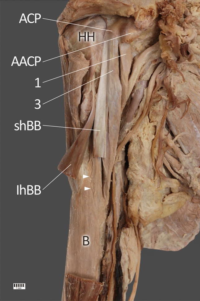
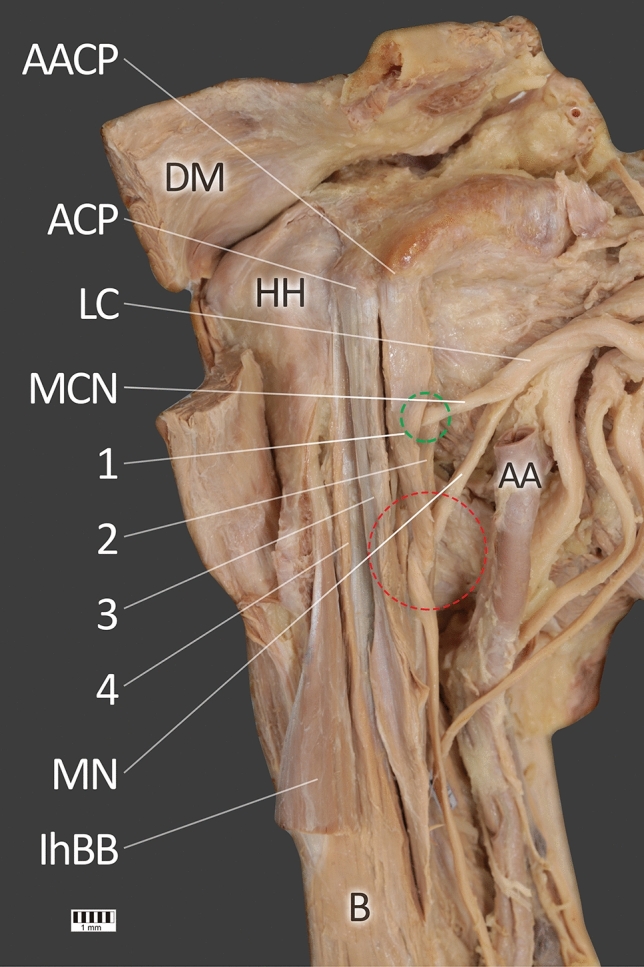
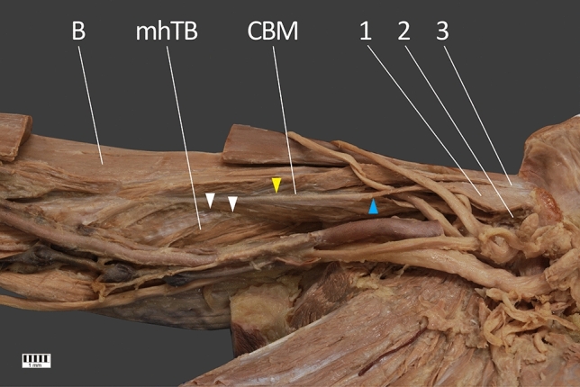
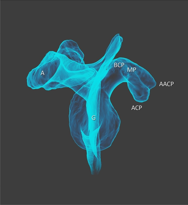
The coracobrachialis muscle (CBM) originates from the apex of the coracoid process, in common with the short head of the biceps brachii muscle, and from the intermuscular septum. Both the proximal and distal attachment of the CBM, as well as its relationship with the musculocutaneus nerve demonstrate morphological variability, some of which can lead to many diseases. The present case study presents a new description of a complex origin type (four-headed CBM), as well as the fusion of both the short biceps brachii head, brachialis muscle and medial head of the triceps brachii. In addition, the first and second heads formed a tunnel for the musculocutaneus and median nerves. This case report has clear clinical value due to the split mature of the coracoid process, and is a significant indicator of the development of interest in this overlooked muscle.
Keywords: Anatomical variations; Coracobrachialis muscle; Median nerve; Musculocutaneus nerve; Split coracoid.
Conflict of interest statement
The authors declare that they have no competing interests.
Figures


Similar articles
-
A proposal for a new classification of coracobrachialis muscle morphology.Surg Radiol Anat. 2021 May;43(5):679-688. doi: 10.1007/s00276-021-02700-1. Epub 2021 Feb 9. Surg Radiol Anat. 2021. PMID: 33564931 Free PMC article.
-
Coracobrachialis muscle variants in human fetuses.Ann Anat. 2025 Feb;258:152372. doi: 10.1016/j.aanat.2024.152372. Epub 2024 Dec 18. Ann Anat. 2025. PMID: 39706309
-
Potential compression of the musculocutaneous, median and ulnar nerves by a very rare variant of the coracobrachialis longus muscle.Folia Morphol (Warsz). 2021;80(3):707-713. doi: 10.5603/FM.a2020.0085. Epub 2020 Aug 26. Folia Morphol (Warsz). 2021. PMID: 32844391
-
A rare accessory coracobrachialis muscle: a review of the literature.Surg Radiol Anat. 2003 Feb;24(6):406-10. doi: 10.1007/s00276-002-0079-5. Epub 2003 Feb 4. Surg Radiol Anat. 2003. PMID: 12652369 Review.
-
A double communication branch between musculocutaneous and median nerves: first case report, anatomical study, and comprehensive review of clinical implications.Eur Rev Med Pharmacol Sci. 2024 Oct;28(19):4376-4382. doi: 10.26355/eurrev_202410_36833. Eur Rev Med Pharmacol Sci. 2024. PMID: 39436082 Review.
Cited by
-
Proportional localisation of the entry point of the coracobrachialis muscle by the musculocutaneous nerve along the humerus.Eur J Trauma Emerg Surg. 2023 Feb;49(1):299-306. doi: 10.1007/s00068-022-02063-1. Epub 2022 Jul 24. Eur J Trauma Emerg Surg. 2023. PMID: 35871667
-
The Subscapularis Muscle: A Proposed Classification System.Biomed Res Int. 2021 Dec 11;2021:7450000. doi: 10.1155/2021/7450000. eCollection 2021. Biomed Res Int. 2021. PMID: 34931169 Free PMC article.
-
Is it the coracobrachialis superior muscle, or is it an unidentified rare variant of coracobrachialis muscle?Surg Radiol Anat. 2021 Oct;43(10):1581-1586. doi: 10.1007/s00276-021-02773-y. Epub 2021 May 26. Surg Radiol Anat. 2021. PMID: 34037825 Free PMC article.
-
Three-Headed Biceps Brachii Muscle: A Rare Site of Proximal Median Nerve Entrapment.Cureus. 2024 Jun 7;16(6):e61886. doi: 10.7759/cureus.61886. eCollection 2024 Jun. Cureus. 2024. PMID: 38975522 Free PMC article.
-
Relationships among Coracobrachialis, Biceps Brachii, and Pectoralis Minor Muscles and Their Correlation with Bifurcated Coracoid Process.Biomed Res Int. 2022 Mar 25;2022:8939359. doi: 10.1155/2022/8939359. eCollection 2022. Biomed Res Int. 2022. PMID: 35419460 Free PMC article.
References
-
- Bechtol CO. Coracobrachialis brevis. Clin Orthop. 1954;4:152. - PubMed
Publication types
MeSH terms
LinkOut - more resources
Full Text Sources
Miscellaneous
