Review
. 2021 Jan;1868(1):118867.
doi: 10.1016/j.bbamcr.2020.118867. Epub 2020 Oct 2.

Getting out what you put in: Copper in mitochondria and its impacts on human disease

Affiliations
Review

Getting out what you put in: Copper in mitochondria and its impacts on human disease

Paul A Cobine et al. Biochim Biophys Acta Mol Cell Res. 2021 Jan.

Abstract

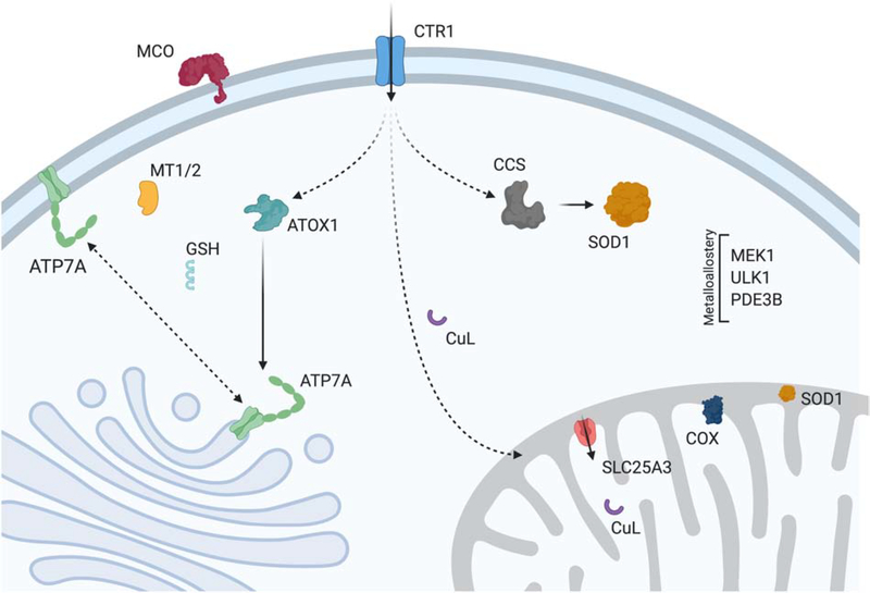
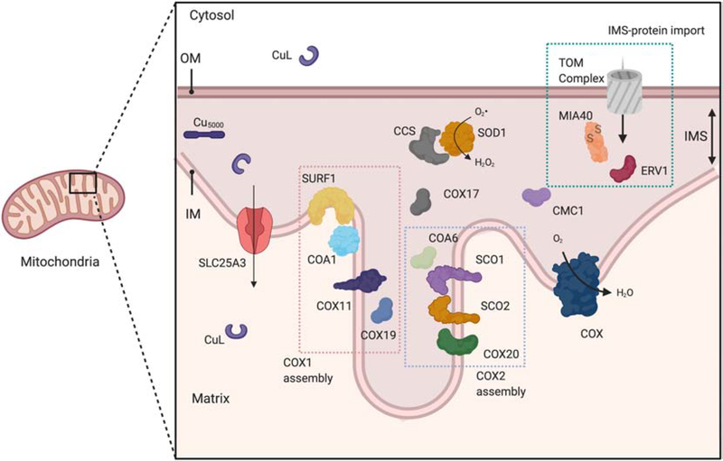
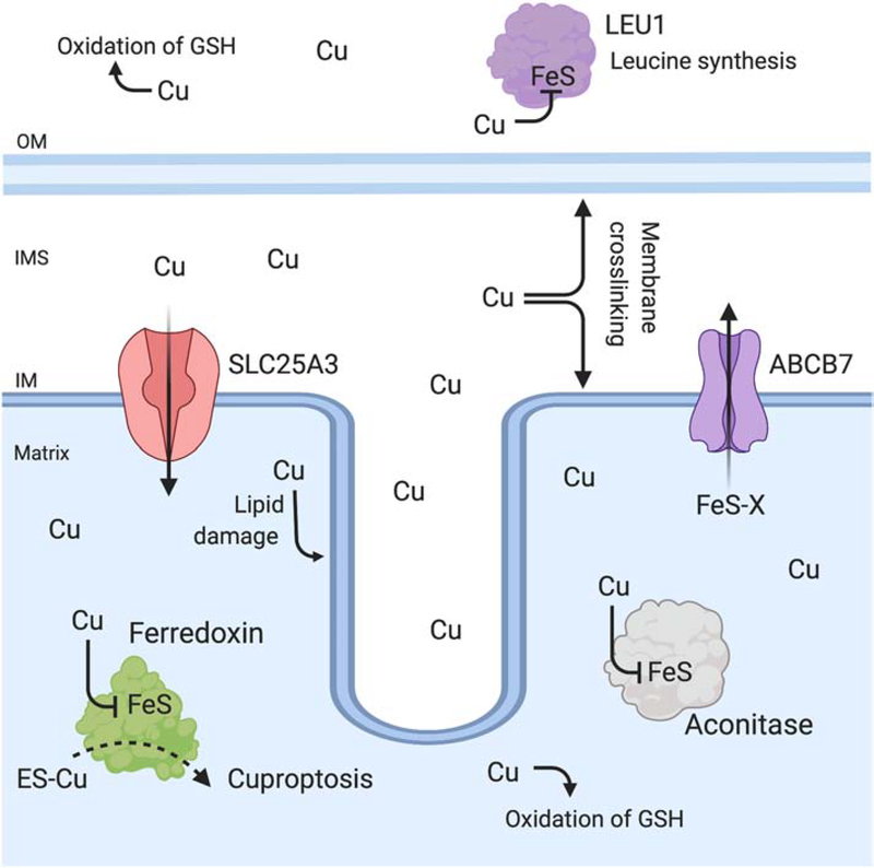
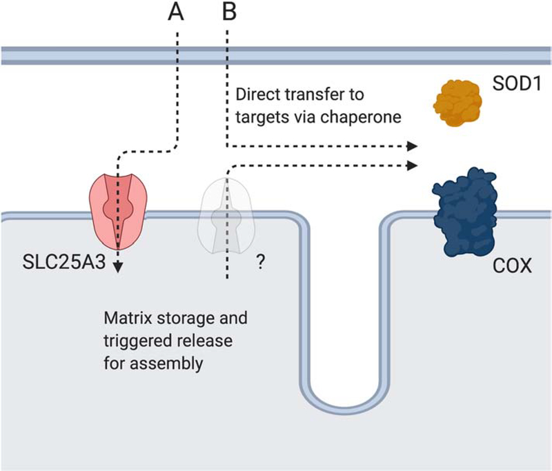
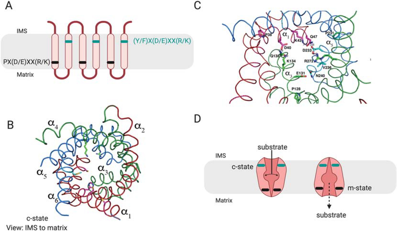
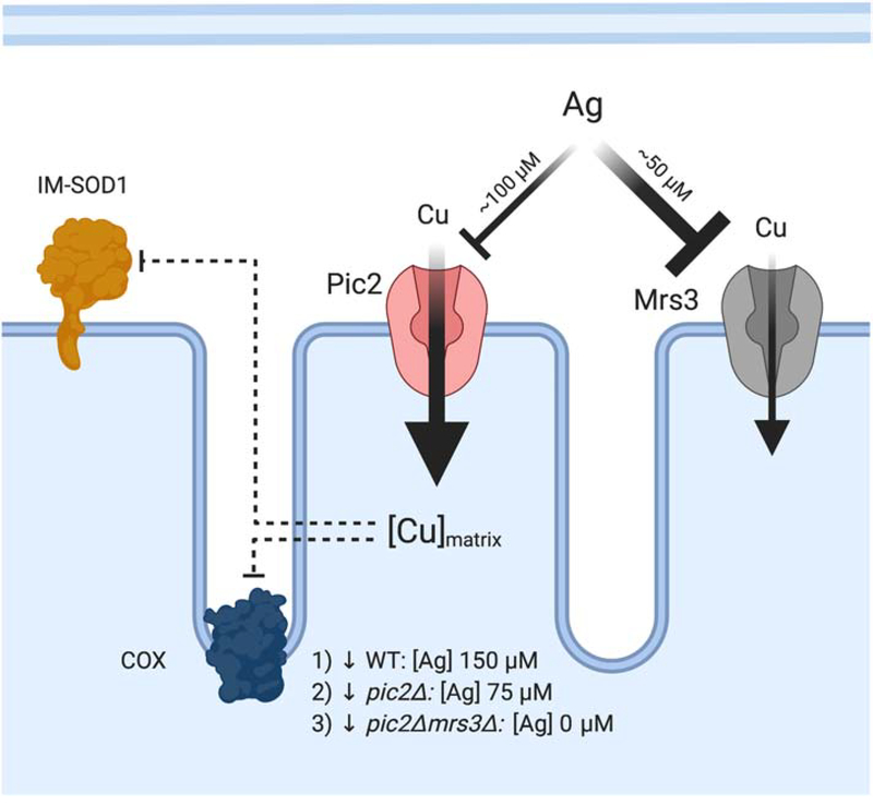
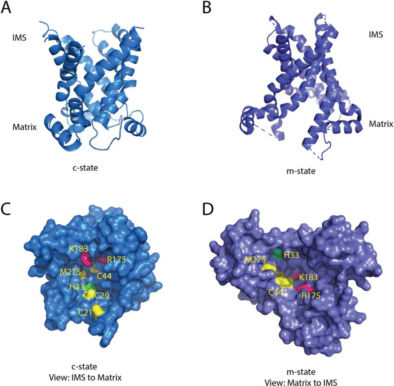
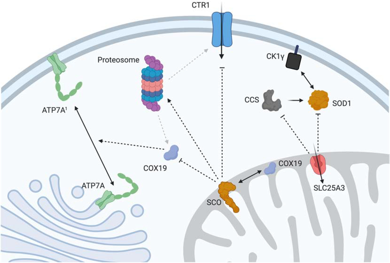
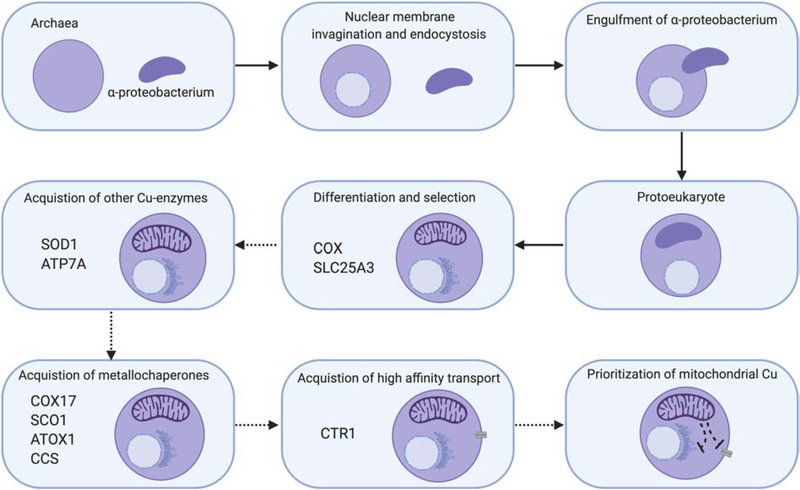
Mitochondria accumulate copper in their matrix for the eventual maturation of the cuproenzymes cytochrome c oxidase and superoxide dismutase. Transport into the matrix is achieved by mitochondrial carrier family (MCF) proteins. The major copper transporting MCF described to date in yeast is Pic2, which imports the metal ion into the matrix. Pic2 is one of ~30 MCFs that move numerous metabolites, nucleotides and co-factors across the inner membrane for use in the matrix. Genetic and biochemical experiments showed that Pic2 is required for cytochrome c oxidase activity under copper stress, and that it is capable of transporting ionic and complexed forms of copper. The Pic2 ortholog SLC25A3, one of 53 mammalian MCFs, functions as both a copper and a phosphate transporter. Depletion of SLC25A3 results in decreased accumulation of copper in the matrix, a cytochrome c oxidase defect and a modulation of cytosolic superoxide dismutase abundance. The regulatory roles for copper and cuproproteins resident to the mitochondrion continue to expand beyond the organelle. Mitochondrial copper chaperones have been linked to the modulation of cellular copper uptake and export and the facilitation of inter-organ communication. Recently, a role for matrix copper has also been proposed in a novel cell death pathway termed cuproptosis. This review will detail our understanding of the maturation of mitochondrial copper enzymes, the roles of mitochondrial signals in regulating cellular copper content, the proposed mechanisms of copper transport into the organelle and explore the evolutionary origins of copper homeostasis pathways.

Keywords: Copper; Cytochrome c oxidase; Mitochondria; Mitochondrial carrier family; Superoxide dismutase.


Figures

Figure 1:
Figure 1:. Cu homeostasis in mammals
Cu enters the cell via the high affinity plasma membrane transporter CTR1 and then is distributed to various targets via metallochaperones (ATOX1, CCS) or alternative ligands (CuL, GSH). Cu enters the trans-Golgi network through an interaction between ATOX1 and ATP7A. Once the Cu enters the cisternae, it can be incorporated into numerous enzymes including the multi-Cu oxidases (MCO). In mitochondria, Cu is transported into the matrix by SLC25A3 for its storage. Upon being triggered by an unknown mechanism Cu is translocated back across the IM to IMS-localized metallochaperones which use it to facilitate assembly of cytochrome c oxidase (COX) and maturation of superoxide dismutase (SOD1). Cu in the cytosol is used as a co-factor by SOD1 for protection against oxidative stress, and CCS is required for metallation of this site and the formation of an essential disulfide bond. When Cu reaches excess levels, it is bound by metallothionein (MT1/2) and ATP7A relocalizes to the PM where it acts as a Cu exporter. In specific cell types, ATP7A is replaced by ATP7B which relocalizes to exocytic vesicles during Cu stress to promote its excretion via the bile (not shown). Dynamic Cu pools within the cell bind at allosteric sites that regulate the activity of the kinases MEK1 and ULK1, and the phosphodiesterase PD3EB in adipocytes.
Figure 2:
Figure 2:. Assembly of COX and SOD1 in mitochondria
Mitochondrial IM and IMS enzymes are assembled as a result of the concerted actions of multiple metallochaperones. COX is assembled in modules that are subsequently combined to yield the mature holoenzyme complex. COX1 which contains the CuB site and heme co-factors is assembled as a large, modular complex that include SURF1, COA1 and COX11. COX19 is involved in the reduction of the Cu-binding cysteines and activation of COX11. The CuA site is assembled by a complex suite of redox-dependent interactions that ultimately allow SCO1 to insert Cu into apo-COX2. COX20 physically interacts with apo-COX2 upon its insertion into the IM, and COA6 and SCO2 work in concert to ensure that the active site cysteines in SCO1 and COX2 are maintained in the reduced state during CuA site maturation. COX17 supplies the Cu necessary to assemble both the COX1 and COX2 modules. CMC1 is also involved in COX assembly, and genetic studies in yeast support an additional role for CMC1 in promoting SOD1 activity. CCS delivers Cu to SOD1 and forms the disulfide bond required for its activity. The Cu used in these reactions is proposed to come from the mitochondrial matrix. The Cu is imported by SLC25A3 after being delivered across the cytosol by an unknown non-proteinaceous ligand (CuL). An alternate hypothesis is that Cu is stored in an IMS complex (Cu5000) that interacts directly with the metallochaperone proteins resident to this compartment. The cysteine containing IMS chaperones (e.g. COA6, CMC1, COX17, COX19, CCS, SOD1) are imported into the IMS via the TOM complex of the outer membrane and the MIA4/ERV1 disulfide relay machinery.
Figure 3:
Figure 3:. Cu toxicity in mitochondria
Cu is essential yet becomes toxic when its content exceeds cellular capacity for binding Cu in inert complexes. In mitochondria, Cu toxicity results in lipid damage, protein oxidation, redox imbalance due to inappropriate binding to cysteine-rich sites and depletion of reduced glutathione (GSH). In addition, Cu disrupts and displaces Fe from exposed FeS cluster containing enzymes. Cu has been shown to inactivate ferredoxin and aconitase, which are both localized to the matrix. Cu also inactivates cytosolic enzymes required for leucine synthesis (LEU1). In fungal models, deletion of the ABCB7-homolog (Atm1) responsible for transporting a FeS intermediate of unknown identity (FeS-X) increases Cu toxicity, and emphasizes that cytosolic targets are also an important aspect of Cu toxicity. The ionophore elesclomol (ES-Cu) increases Cu accumulation in mitochondria, and at high concentrations induces cell death via a ferredoxin-dependent mechanism named cuproptosis.
Figure 4:
Figure 4:. Structure of COX and SOD1
A) The intact, 14-subunit structure of human cytochrome c oxidase (PDB: 5z62) represented as a cartoon backbone with the 3 mitochondrially-encoded subunits highlighted in blue. B) COX structure with the mitochondrial subunits removed to reveal the heme and Cu co-factors. C) Enlarged rendering of the bi-nuclear CuA site, the heme a and the mixed metal heme a3-CuB site. D) Cartoon depiction of one half of the SOD1 dimer (PDB: 2c9v) showing its Cu and zinc co-factors.
Figure 5:
Figure 5:. Cu recruitment from the cytosol for COX and IMS-SOD1 maturation
Two alternative pathways may exist for Cu delivery to COX and IMS-SOD1. Pathway A depicts the matrix storage pathway where Cu that was recruited to the matrix by SLC25A3 for storage is transported back to the IMS by an unidentified transporter for enzyme maturation. The existence of this pathway is supported by multiple studies under Cu stress. Pathway B depicts the direct access pathway where chaperones bind Cu as it enters the IMS or access it after storage in this compartment to support enzyme maturation. Storage in the IMS could be facilitated by the Cu5000 complex that was originally isolated from brain mitochondria.
Figure 6:
Figure 6:. Schematic of MCF structure
MCF proteins have a conserved, repeated three TM helices structure. A) Within the odd numbered helices a PX(D/E)XX(R/K) motif, shown in black, forms a matrix salt bridge that prevents entry of substrates to the matrix and blocks proton leak. A complementary (Y/F)X(D/E)XX(R/K) motif shown in teal is found in the even numbered helices, and forms salt bridges on the IMS side of the protein. B) The cartoon backbone of the modelled structure of yeast Pic2 in the c-state based on the structure of the ADP/ATP exchanger (PDB: 4C9G). The conserved repeated structure is highlighted in red for helices 1 and 2 (α1, α2), green for helices 3 and 4 (α3, α4), blue for helices 5 and 6 (α5, α6). The structure is shown from the IMS side. C) The salt bridge interactions poise the MCF in either the c-state open to the IMS, or the m-state open to the matrix. The transition between these two states requires substrate binding, and the relative strength of the salt bridge interactions determines whether the transporter acts as a uniporter or as an exchanger. Strong IMS and matrix salt bridges would require exchange of a substrate to reset to the opposite state. Any weaker salt bridges would allow the transporter to be unidirectional.
Figure 7:
Figure 7:. Phenotypic analysis of mitochondrial Cu transporters
Cu-transporting MCF proteins were identified using genetic screens based on limiting Cu availability with the addition of Ag as a competitor. At concentrations of 150 μM, Ag inhibits Cu uptake and results in decreased COX activity. To elicit the same defect in cells lacking PIC2 only 75–100 μM Ag is required, suggesting a loss of high-affinity transport. Mrs3 was identified as a lower affinity Cu transporter based on additive phenotypes. Yeast cells lacking PIC2 and MRS3 grown under mild Cu chelation without the addition of Ag had decreased COX, decreased IM-SOD1 activity and lower mitochondrial Cu uptake and accumulation.
Figure 8:
Figure 8:. Structural model of Pic2
A) A cartoon backbone representation of Pic2 modeled onto the c-state of the ADP/ATP exchanger (PDB:4C9G) B) A cartoon backbone representation of Pic2 modeled onto the m-state of the ADP/ATP exchanger (PDB: 6GCI). C) Surface rendering of the Pic2 model in the c-state model looking down the channel from the IMS side of the IM highlighting the positioning of Cys21, Cys29, Cys44, His33, Met275, Arg175 and Lys183 residues in yellow, green and pink. D) Surface rendering of the Pic2 model in the m-state model highlighting the residues listed in C).
Figure 9:
Figure 9:. Mitochondrial Cu-associated regulation of cellular physiology
Genetic and pharmacological manipulation of mitochondrial Cu in mammalian models results in dramatic remodeling of cellular Cu homeostasis. SCO proteins generate a redox signal that is transduced to the cytosol in part by COX19 to trigger the relocalization of a fraction of the ATP7A pool to the PM to facilitate Cu export. In addition, the SCO-mediated signal(s) affects Cu import via CTR1 by promoting its proteasomal degradation or preventing its localization to the PM. While the combination of these two events causes SCO mutant cells to be profoundly Cu deficient, their mitochondrial Cu pool is preserved. In contrast, deletion of SLC25A3 causes a mitochondrial Cu defect that triggers a signal that results in decreased CCS levels and cytosolic SOD1 activity. We speculate that this would affect the known interaction between SOD1 and casein kinase 1γ (CK1γ), thereby regulating glucose utilization in this mutant cell line.
Figure 10:
Figure 10:. Proposed model for Cu homeostasis during eukaryogenesis
Eukaryotes are thought to have evolved from ancient archaea that had initially differentiated to gain a nuclear membrane and acquire endocytic machinery. Thereafter, the endocytosis of an α-proteobacterium started a symbiotic relationship between the two organisms that resulted in permanent innovation, with retention of the α-proteobacterium as a mitochondrion and differentiation into a multi-organelle protoeukaryote. During its adaptation and development, we speculate that COX was one of the major Cu requirements for the protoeukaryote given its role as a co-factor in the primitive enzyme. As its evolution continued, numerous intermediate steps selected for other Cu-requiring processes and the metallochaperones required to enhance their activity and Cu recruitment. The exact order of gains and losses has not been investigated and therefore is shown here with dashed arrows. One of the final steps based on preliminary investigation of diverse genomes from the tree of life was the acquisition of a high affinity Cu transport system at the plasma membrane to fulfill these Cu needs. We propose that to maintain prioritization of Cu for mitochondria, the protoeukaryote then refined the hierarchical regulation of Cu handling around signals originating from the organelle.

Similar articles

Cited by

References

    1. Robinson NJ, Winge DR, Copper metallochaperones, Annu Rev Biochem 79 (2010) 537–62. - PMC - PubMed
    1. Chang CJ, Searching for harmony in transition-metal signaling, Nat Chem Biol 11(10) (2015) 744–7. - PubMed
    1. Krishnamoorthy L, Cotruvo JA Jr., Chan J, Kaluarachchi H, Muchenditsi A, Pendyala VS, Jia S, Aron AT, Ackerman CM, Wal MN, Guan T, Smaga LP, Farhi SL, New EJ, Lutsenko S, Chang CJ, Copper regulates cyclic-AMP-dependent lipolysis, Nat Chem Biol 12(8) (2016) 586–92. - PMC - PubMed
    1. Brady DC, Crowe MS, Turski ML, Hobbs GA, Yao X, Chaikuad A, Knapp S, Xiao K, Campbell SL, Thiele DJ, Counter CM, Copper is required for oncogenic BRAF signalling and tumorigenesis, Nature 509(7501) (2014) 492–6. - PMC - PubMed
    1. Tsang T, Posimo JM, Gudiel AA, Cicchini M, Feldser DM, Brady DC, Copper is an essential regulator of the autophagic kinases ULK1/2 to drive lung adenocarcinoma, Nat Cell Biol 22(4) (2020) 412–424. - PMC - PubMed

Publication types

MeSH terms