

Aloe extract inhibits porcine epidemic diarrhea virus in vitro and in vivo
- PMID: 32979750
- PMCID: PMC7491386
- DOI: 10.1016/j.vetmic.2020.108849
Aloe extract inhibits porcine epidemic diarrhea virus in vitro and in vivo
Abstract
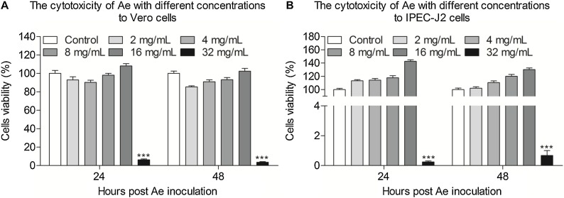
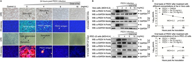
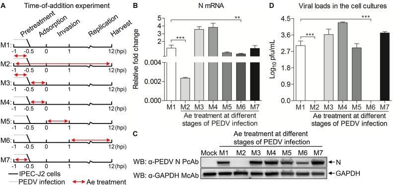
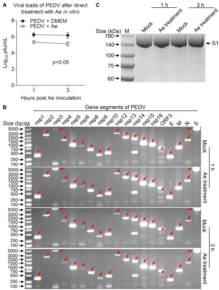
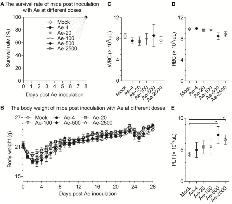
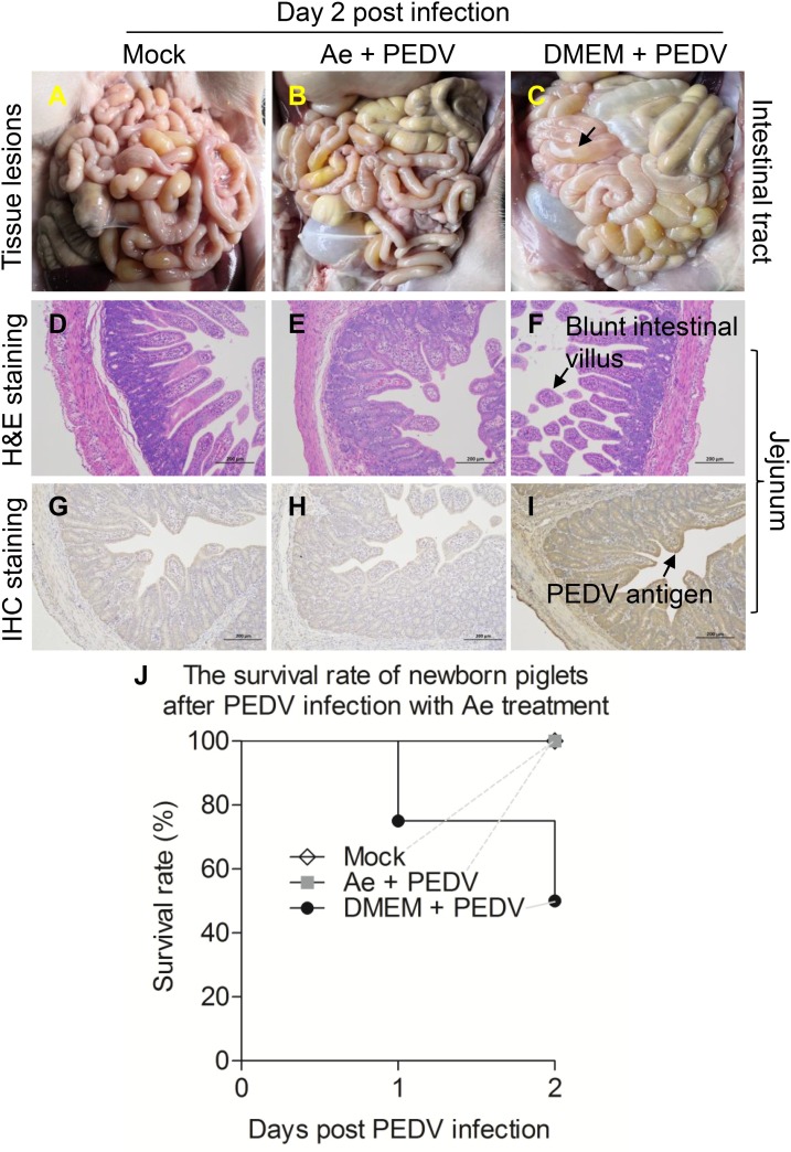
Porcine epidemic diarrhea virus (PEDV) causes severe diarrhoea and high mortality in neonatal suckling piglets, leading to significant economic losses to the swine industry. Currently there are no adequate control strategies against circulating PEDV variants, making an urgent need to exploit effect antiviral therapies to compensate for vaccines. Here, we report that Aloe extract can hamper completely the proliferation of PEDV at a non-cytotoxic concentration of 16 mg/mL determined by CCK-8 assay in Vero and IPEC-J2 cells in vitro. Furthermore, time course analysis indicated the extract exerted its inhibition at the late stage of the viral life cycle. Moreover, we also confirmed that the extract can inactivated PEDV directly but did not act on the viral genome and S1 protein. Importantly, the extract at a relatively safety concentration of 100 mg/kg of body weight, which was confirmed in mice, could reduce virus load and pathological change in intestinal tract of pigs and protect newborn piglets from lethal challenge with highly pathogenic PEDV variant GDS01 infection, indicating that Aloe extract efficiently inhibited PEDV infection in vivo. Collectively, our findings suggest that the aqueous extract from the Aloe could inhibit PEDV replication in vitro and in vivo and might be a good target for drug development against PEDV.
Keywords: Aloe extract; Antiviral activity; Porcine epidemic diarrhea virus (PEDV); Swine.
Copyright © 2020. Published by Elsevier B.V.
Conflict of interest statement
The authors declare that they have no conflict interest.
Figures
Similar articles
-
Advances research in porcine enteric coronavirus therapies and antiviral drugs.Vet Q. 2024 Dec;44(1):1-49. doi: 10.1080/01652176.2024.2421299. Epub 2024 Nov 1. Vet Q. 2024. PMID: 39484691 Free PMC article. Review.
-
Lizhong decoction inhibits porcine epidemic diarrhea virus in vitro and in vivo.J Ethnopharmacol. 2024 Oct 28;333:118428. doi: 10.1016/j.jep.2024.118428. Epub 2024 Jun 7. J Ethnopharmacol. 2024. PMID: 38852639
-
Berbamine inhibits porcine epidemic diarrhea virus in vitro and in vivo.Vet Microbiol. 2024 Nov;298:110244. doi: 10.1016/j.vetmic.2024.110244. Epub 2024 Aug 30. Vet Microbiol. 2024. PMID: 39236425
-
Antiviral effect of lithium chloride on porcine epidemic diarrhea virus in vitro.Res Vet Sci. 2018 Jun;118:288-294. doi: 10.1016/j.rvsc.2018.03.002. Epub 2018 Mar 5. Res Vet Sci. 2018. PMID: 29547727 Free PMC article.
-
Traditional Chinese medicine as a promising choice for future control of PEDV.Virus Res. 2025 Jun;356:199572. doi: 10.1016/j.virusres.2025.199572. Epub 2025 Apr 10. Virus Res. 2025. PMID: 40220931 Review.
Cited by
-
Bis-Benzylisoquinoline Alkaloids Inhibit Porcine Epidemic Diarrhea Virus In Vitro and In Vivo.Viruses. 2022 Jun 6;14(6):1231. doi: 10.3390/v14061231. Viruses. 2022. PMID: 35746702 Free PMC article.
-
NS7a of SADS-CoV promotes viral infection via inducing apoptosis to suppress type III interferon production.J Virol. 2024 May 14;98(5):e0031724. doi: 10.1128/jvi.00317-24. Epub 2024 Apr 16. J Virol. 2024. PMID: 38624231 Free PMC article.
-
Advances research in porcine enteric coronavirus therapies and antiviral drugs.Vet Q. 2024 Dec;44(1):1-49. doi: 10.1080/01652176.2024.2421299. Epub 2024 Nov 1. Vet Q. 2024. PMID: 39484691 Free PMC article. Review.
-
Utilization of Aloe Compounds in Combatting Viral Diseases.Pharmaceuticals (Basel). 2022 May 13;15(5):599. doi: 10.3390/ph15050599. Pharmaceuticals (Basel). 2022. PMID: 35631425 Free PMC article. Review.
-
In vitro suppression of porcine epidemic diarrhea virus by Panax notoginseng saponins: assessing antiviral potential.Arch Virol. 2024 Apr 2;169(5):89. doi: 10.1007/s00705-024-06020-8. Arch Virol. 2024. PMID: 38565720
References
-
- Birgit Quinting, Béatrice Robert, Carine Letellier, Mathieu Boxus, Pierre Kerkhofs. Development of a 1-step enzyme-linked immunosorbent assay for the rapid diagnosis of bovine respiratory syncytial virus in postmortem specimens. J. Vet. Diagn. Invest. 2007;19:238–243. - PubMed
-
- Chen D., Xiong Y., Wang L., Lv B., Lin Y. Characteristics of emodin on modulating the contractility of jejunal smooth muscle. Can. J. Physiol. Pharmacol. 2012;90:455–462. - PubMed
-
- Debouck P., Pensaert M.B. Experimental infection of pigs with a new porcine enteric coronavirus, CV 777. Am. J. Vet. Res. 1980;41:219–223. - PubMed
MeSH terms
Substances
LinkOut - more resources
Full Text Sources
