. 2020 Oct:60:103014.
doi: 10.1016/j.ebiom.2020.103014. Epub 2020 Sep 24.

Hypothermic oxygenated perfusion protects from mitochondrial injury before liver transplantation

Affiliations

Hypothermic oxygenated perfusion protects from mitochondrial injury before liver transplantation

Andrea Schlegel et al. EBioMedicine. 2020 Oct.

Abstract

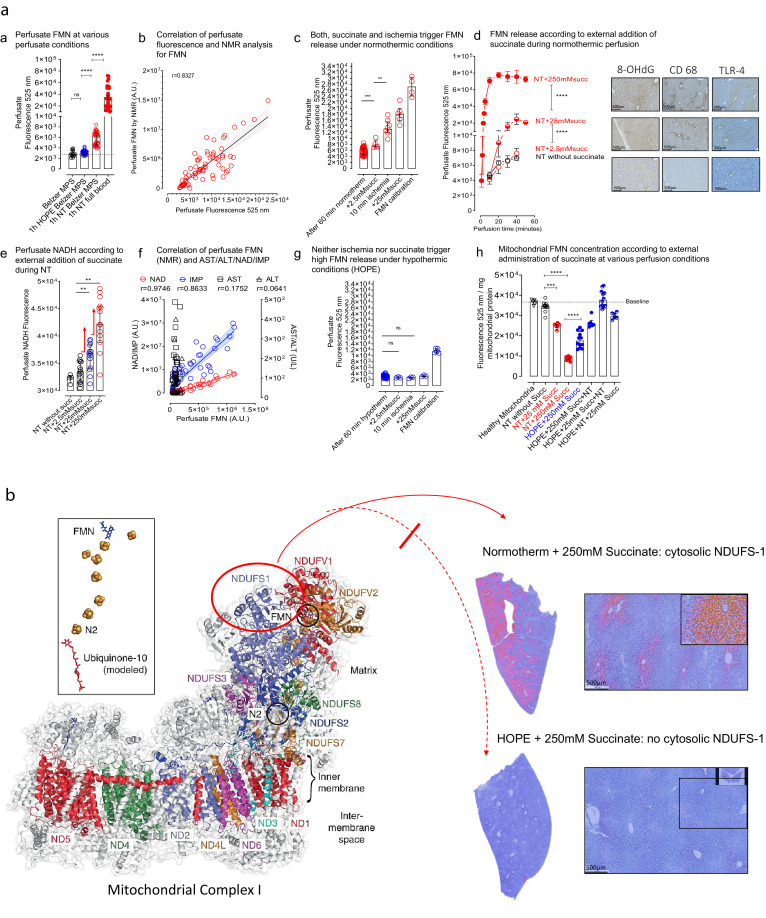
Background: Mitochondrial succinate accumulation has been suggested as key event for ischemia reperfusion injury in mice. No specific data are however available on behavior of liver mitochondria during ex situ machine perfusion in clinical transplant models.

Methods: We investigated mitochondrial metabolism of isolated perfused rat livers before transplantation. Livers were exposed to warm and cold ischemia to simulate donation after circulatory death (DCD) and organ transport. Subsequently, livers were perfused with oxygenated Belzer-MPS for 1h, at hypothermic or normothermic conditions. Various experiments were performed with supplemented succinate and/or mitochondrial inhibitors. The perfusate, liver tissues, and isolated mitochondria were analyzed by mass-spectroscopy and fluorimetry. Additionally, rat DCD livers were transplanted after 1h hypothermic or normothermic oxygenated perfusion. In parallel, perfusate samples were analysed during HOPE-treatment of human DCD livers before transplantation.

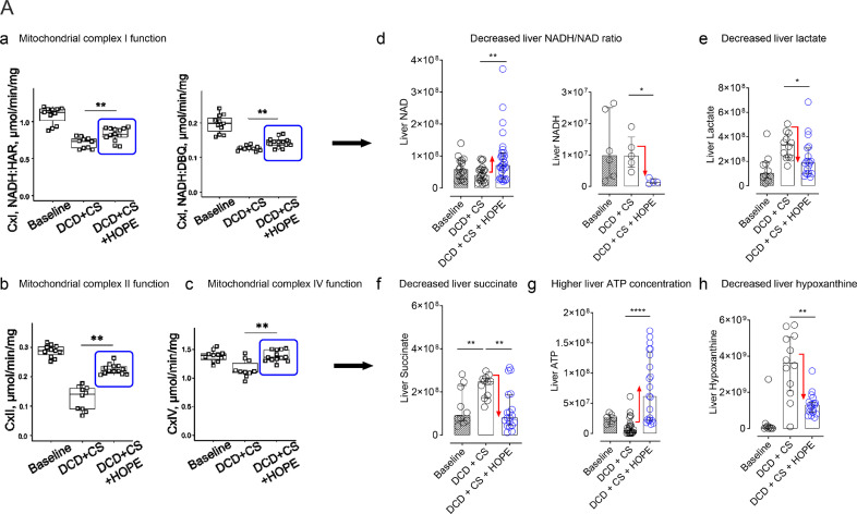
Findings: Succinate exposure during rat liver perfusion triggered a dose-dependent release of mitochondrial Flavin-Mononucleotide (FMN) and NADH in perfusates under normothermic conditions. In contrast, perfusate FMN was 3-8 fold lower under hypothermic conditions, suggesting less mitochondrial injury during cold re-oxygenation compared to normothermic conditions. HOPE-treatment induced a mitochondrial reprogramming with uploading of the nucleotide pool and effective succinate metabolism. This resulted in a clear superiority after liver transplantation compared to normothermic perfusion. Finally, the degree of mitochondrial injury during HOPE of human DCD livers, quantified by perfusate FMN and NADH, was predictive for liver function.

Interpretation: Mitochondrial injury determines outcome of transplanted rodent and human livers. Hypothermic oxygenated perfusion improves mitochondrial function, and allows viability assessment of liver grafts before implantation.

Funding: detailed information can be found in Acknowledgments.

Keywords: Complex I; FMN; Hypothermic oxygenated perfusion; Liver transplantation; Normothermic oxygenated perfusion.


Conflict of interest statement

Declaration of Interests This study was conducted at the University Hospital Zurich (USZ). Raw data and laboratory analysis results were extracted at the laboratory of the Department of Visceral Surgery and Transplantation of the USZ. Data analysis was carried out at the laboratory of the Department of Visceral Surgery and Transplantation of the USZ. The authors have declared that no competing interests exist. The study was principally funded by a main research grant from the swiss national foundation (SNF), awarded to Professor Philipp Dutkowski (Ref: 32003B-140776/1, 3200B-153012/1, 31IC30-166909). Mitochondrial and tissue analysis was further supported by the Max Planck Society (Dr. David Meierhofer) and the NIH grant: R01NS112381 awarded to Dr. Alexander Galkin. There are no patents involved or affecting this study.

Figures

Image, graphical abstract
Graphical abstract
Fig 1:
Fig. 1
Perfusion circuit and online fluorescence of FMN The perfusion circuit used for all rat liver perfusions included a temperature control device, flow and pressure sensors, the perfusion chamber, where the liver was placed with another temperature sensor, the sampling port, an injection port for the application of succinate or mitochondrial blockers, and a flow cell at the outflow of the liver to quantify the fluorescence (a). Detection of the emitted light was quantified between 500–600 nm, while excitation was performed at a wavelength of 450 nm. Normo- or hypothermic oxygenated perfusion was performed with a computer adjusted pressure-controlled system to automatically limit perfusion pressure at 12 mm Hg (normothermic) or at 3 mm Hg (hypothermic), and to allow the liver to autoregulate the flow, which becomes subsequently detected by a flow sensor. Real time response with an increasing fluorescence is demonstrated in accordance with succinate supplementation during normothermic perfusion, while under hypothermic oxygenated conditions, much lower perfusate fluorescence FMN release was detected despite succinate trigger (b).
Fig 2
Fig. 2
a: Detection of mitochondrial FMN and metabolite release in perfusates: Release of FMN occurred at various perfusate conditions, including hypothermic or normothermic acellular perfusion, and by normothermic perfusion with whole blood, which resulted in highest FMN release (n = 20) (a). Perfusate FMN, detected by fluorescence, was validated by NMR analysis (Rho = 0.8327) (measured in all rodent samples n > 55) (b). Administration of succinate or intermittent ischemia/reperfusion triggered relevant FMN release during normothermic perfusion. (n = 6–7 for all groups other than: 60 min normotherm alone: n = 19 and n = 4 in FMN calibration) (c). Release of FMN increased in accordance with the amount of administered succinate (2.5 mM < 25 mM < 250 mM succinate; n = 6–7 for all groups) (d). Perfusate NADH quantification through spectroscopy demonstrated increasing concentrations according to administered succinate (n = 15 for all groups other than: NT without succinate, n = 5) (e). Perfusate FMN concentrations correlated also well with NAD (Rho = 0.9746) and IMP (Rho = 0.8633), in contrast to release of cytosolic liver enzymes (AST, ALT) (n = 50, human samples) (f). Neither succinate application or intermittent ischemia/reperfusion was found to trigger high FMN release during hypothermic oxygenated perfusion (n = 6 other than calibration group, n = 4 and end of 60 min hypotherm: n = 18) (g). Mitochondrial FMN concentrations were inversely correlated to perfusate FMN, with low intramitochondrial FMN after normothermic perfusion. In contrast, mitochondria were significantly protected from FMN loss during hypothermic oxygenated perfusion despite succinate application (n = 6–12) (h) (Mann-Whitney U-test was used for statistical analysis). Fig. 2b: Imaging of mitochondrial complex I injury: cytosolic staining of released NDUFS-1: With high liver injury, NDUFS-1, a subunit of complex I dissociates from complex 1 and is released into the cytosol. Liver tissues were stained for NDUFS-1 in different groups. Normothermic perfusion with 250 mM Succinate led to high injuries of the mitochondrial complex I as shown by a high number of NDUFS-1 positive hepatocytes in the staining, when compared to hypothermic perfusion, where almost no NDUFS-1-positive cytosols were seen.
Fig 3:
Fig. 3
Downstream inflammation under conditions with maximal succinate administration in perfusates: Histological images of various cellular compounds in livers perfused under normothermic or hypothermic conditions with exposure to perfusate succinate. Parenchymal and non-parenchymal liver cells were protected from oxidative injury and activation under hypothermic conditions. In contrast, reperfusion under normothermic conditions induced oxidative injury, toll like receptor- and Kupffer cell activation (n = 6 and 8, and 2 HPF/experiment) (a). These findings were paralleled by higher perfusate NADH concentrations (n = 15, duplicates) (b), higher oxidated DNA (n = 6 and 8) (C), and higher release of danger signals (HMGB-1) during normothermic perfusion, when compared to hypothermic conditions (n = 6) (d) (Mann-Whitney U-test was used for statistical analysis).
Fig 4:
Fig. 4
Assessment of mitochondrial function during normo- and hypothermic perfusion in the presence of mitochondrial inhibitors: Various mitochondrial inhibitors were administered during liver perfusion together with succinate. Inhibition of complex II by malonate triggered increase of perfusate NADH during normothermic (a) and during hypothermic conditions (b). However, complex II inhibition caused only under normothermic conditions relevant FMN release (c), in contrast to hypothermic perfusion (HOPE) (d). During HOPE, high FMN release occurred with a combination of complex I and complex II inhibitors, or by excess of NADH donors (d). Administration of increasing succinate, or administration of a combination of succinate and malonate, led to increasing NAD in perfusates under normothermic conditions (e). Succinate plus malonate resulted also in increased purine salvage, detectable by high IMP (f) simultaneously with low hypoxanthine and xanthine (n =8–10, single measurements or duplicates) (g, h) (Mann-Whitney U-test was used for statistical analysis).
Fig 5:
Fig. 5
Mechanism of reoxygenation under normo- and hypothermic conditions: Under conditions of normothermic reperfusion in the presence of increased intra-mitochondrial succinate concentrations ①, complex I-bound FMN dissociates due to over-reduction of NADH dehydrogenase②. A reductive dissociation of FMNH2 would be followed by a rapid oxidation to FMN by oxygen with ROS formation③ and significant mitochondrial dysfunction, leading to ATP breakdown ④. As a consequence, mitochondrial oxidative injury occurs under normothermic reperfusion conditions, and is further aggravated by malonate (a). In contrast, FMN release and oxidative stress decreases significantly under hypothermic reperfusion conditions, despite succinate provoke and despite the presence of malonate (b).
Fig 6
Fig. 6
a: Mitochondrial complex I–IV function without and with hypothermic oxygenated perfusion (HOPE): Hypothermic oxygenated perfusion (HOPE), applied in DCD livers after cold ischemia, improved complex I, II and IV function (n = 11–12)(a, b, c). Subsequently, this resulted in decreased NADH/NAD tissue ratios (d), while lactate and succinate were metabolized (e, f), and liver ATP was uploaded (g) by purine salvage pathways (h). Fig. 6b: Mitochondrial reprogramming by hypothermic oxygenated perfusion (HOPE): Isolated mitochondria were analyzed by metabolite extraction and targeted liquid chromatography mass spectroscopy in four experimental groups, e.g. in normothermic perfusion (NT) of rat DCD livers, without or with high perfusate succinate (250 mM), and in HOPE treated rat DCD livers, with application of 250 mM succinate already in the HOPE perfusate, followed by normothermic perfusion. Despite the presence of high intramitochondrial succinate load during HOPE treatment, mitochondria maintained functioning with preserved FMN and NAD, metabolized succinate, and uploaded purine nucleotides, in contrast to untreated livers (n = 10–12, except NADH: n = 6) (Mann-Whitney U-test was used for statistical analysis). Fig. 6c: DCD liver injury & ATP 24 h after liver transplantation: Significantly lower liver injury was found after transplantation of HOPE treated livers compared to normothermic perfusion, as shown by liver transaminases, measured in the recipient 24 h after DCD liver transplantation (n = 5–7) (a). Such findings were paralleled by higher cellular energy after HOPE treatment, when compared to normothermic perfusion, measured through ATP content (n = 6–8) (b). The liver histology confirmed these findings with more signs of injury after normothermic perfusion and transplantation, compared to HOPE (n = 6, duplicates of 2HPF per experiment per group) (c) (Mann-Whitney U-test was used for statistical analysis).
Fig 7:
Fig. 7
Human perfusate analysis during HOPE: Human livers donated after circulatory death (DCD), or donated after brain death (DBD), underwent hypothermic oxygenated perfusion (HOPE) for 1–2h after cold storage prior to liver implantation. Perfusate fluorescence was measured during HOPE in 50 livers. Corresponding to experimental data in rodents, human livers released FMN during HOPE (a). The amount of perfusate FMN within the first 30 min correlated well with graft outcome after transplantation, with a discriminative threshold of 8 × 103 arbitrary units of perfusate FMN fluorescence, e.g. 100 ng FMN/ml circulating perfusate = 0.2 μg FMN release/g liver (3l perfusate, 1.5 kg liver). Accordingly, high FMN perfusate concentrations resulted in a very high percentage of graft loss after transplantation (6/9, 67%), in contrast to good outcome at low FMN perfusate values (1/41, 2.4 %) (a). FMN release during HOPE also discriminated highly between DCD and DBD livers (b), and FMN quantification at 30 min of hypothermic oxygenated perfusion revealed an excellent c-statistic (0.9441) by ROC curve analysis (c). Mitochondrial metabolites were further quantified in the HOPE perfusate of human livers. The highest fold increase, comparing lost vs survived grafts, was found for perfusate NAD and FMN (d). Predictive discrimination was achieved through either quantification of perfusate FMN within the first 30 min (area under curve, AUC 1-30 min) or for single perfusate FMN measurements at 30 min (n = 50 human livers) (e). NADH quantification at 30 min of HOPE treatment in human livers confirmed similar significant differences between lost and survived grafts after liver transplantation (7 grafts were lost, measurement done in triplicates) (f) (Mann-Whitney U-test was used for statistical analysis). Perfusate levels of human DCD livers with high injury (graft loss) were in the range of rat livers perfusions with high doses of succinate, underlining the clinical relevance of the model (g).

References

    1. Murphy MP. How mitochondria produce reactive oxygen species. Biochem J [Internet] 2009;417(1):1–13. http://www.ncbi.nlm.nih.gov/pubmed/19061483%5Cnhttp://www.pubmedcentral.... Available from: - DOI - PMC - PubMed
    1. Kim M, Stepanova A, Niatsetskaya Z, Sosunov S, Arndt S, Murphy MP. Attenuation of oxidative damage by targeting mitochondrial complex I in neonatal hypoxic-ischemic brain injury. Free Radic Biol Med. 2018 - PMC - PubMed
    1. Chouchani ET, Pell VR, Gaude E, Aksentijević D, Sundier SY, Robb EL. Ischaemic accumulation of succinate controls reperfusion injury through mitochondrial ROS. Nature [Internet] 2014;515(V):431–435. doi: 10.1038/nature13909. Available from: - DOI - PMC - PubMed
    1. Van Raemdonck D, Neyrinck A, Rega F, Devos T, Pirenne J. Machine perfusion in organ transplantation: a tool for ex-vivo graft conditioning with mesenchymal stem cells? Curr Opin Organ Transpl. 2013;18(1):24–33. - PubMed
    1. Nasralla D, Coussios CC, Mergental H, Akhtar MZ, Butler AJ, Ceresa CDL. A randomized trial of normothermic preservation in liver transplantation. Nature [Internet] 2018 http://www.nature.com/articles/s41586-018-0047-9 Available from: - PubMed

MeSH terms