Review
. 2021 Mar;45(3):205-221.
doi: 10.1111/aor.13827. Epub 2020 Nov 4.

Advances in extracorporeal membrane oxygenator design for artificial placenta technology

Affiliations
Review

Advances in extracorporeal membrane oxygenator design for artificial placenta technology

David G Blauvelt et al. Artif Organs. 2021 Mar.

Abstract

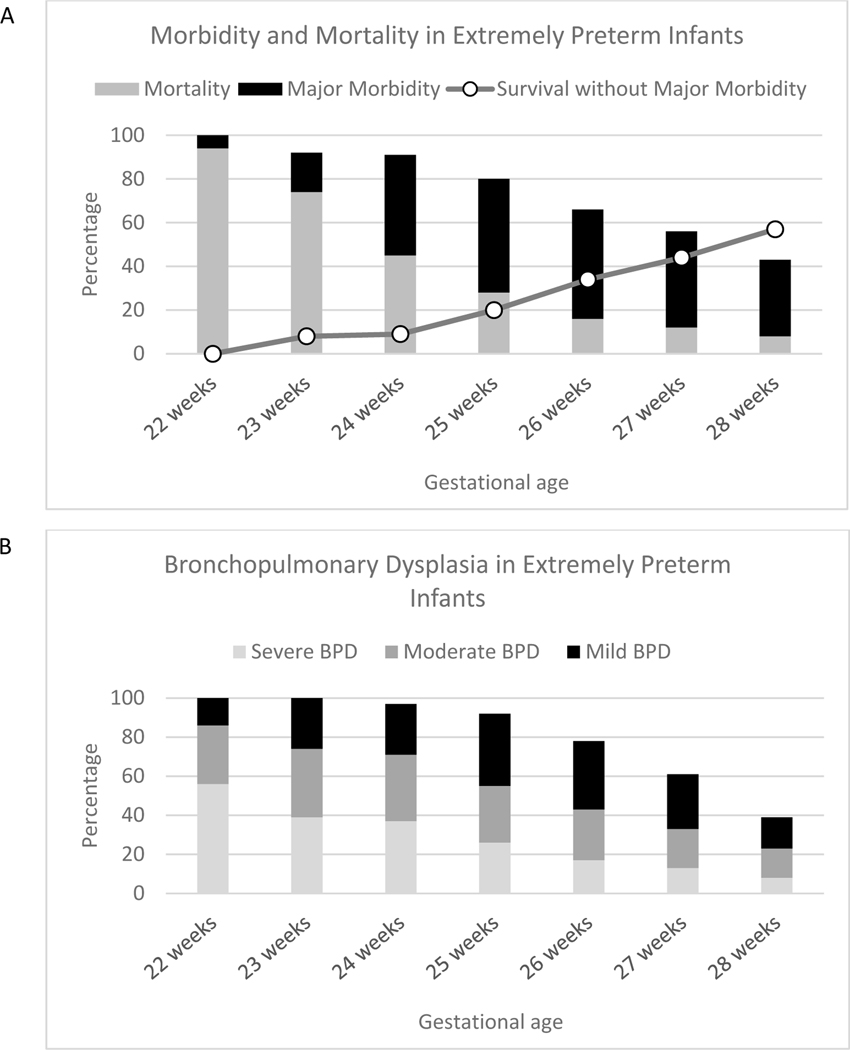
Extreme prematurity, defined as a gestational age of fewer than 28 weeks, is a significant health problem worldwide. It carries a high burden of mortality and morbidity, in large part due to the immaturity of the lungs at this stage of development. The standard of care for these patients includes support with mechanical ventilation, which exacerbates lung pathology. Extracorporeal life support (ECLS), also called artificial placenta technology when applied to extremely preterm (EPT) infants, offers an intriguing solution. ECLS involves providing gas exchange via an extracorporeal device, thereby doing the work of the lungs and allowing them to develop without being subjected to injurious mechanical ventilation. While ECLS has been successfully used in respiratory failure in full-term neonates, children, and adults, it has not been applied effectively to the EPT patient population. In this review, we discuss the unique aspects of EPT infants and the challenges of applying ECLS to these patients. In addition, we review recent progress in artificial placenta technology development. We then offer analysis on design considerations for successful engineering of a membrane oxygenator for an artificial placenta circuit. Finally, we examine next-generation oxygenators that might advance the development of artificial placenta devices.

Keywords: artificial placenta; extracorporeal life support; extreme prematurity; hollow-fiber membranes; microfluidics.


Conflict of interest statement

Conflicts of Interest:

The authors report no conflicts of interest.

Figures

Figure 1:
Figure 1:
Outcomes in extremely premature infants. (a) Mortality and major morbidity rates are high in EPT infants. Survival without major morbidity is correlated with gestational age, with the worst outcomes seen in the most premature infants. Major morbidity is defined as severe intraventricular hemorrhage (IVH), periventricular leukomalacia (PVL), bronchopulmonary dysplasia (BPD), necrotizing enterocolitis (NEC), infections, and stage 3 retinopathy of prematurity (ROP). (b) Bronchopulmonary dysplasia is common in EPT infants and is correlated with gestational age. Both the frequency and severity of BPD increase with decreasing gestational age. Graphs created using data from Stoll et al.
Figure 2:
Figure 2:
Circuit design of a neonatal ECLS circuit. Deoxygenated blood is drawn from a large vein, often in the neck. It is then pumped to an oxygenator, where a blenderized combination of oxygen and compressed air mixes with the blood via a semipermeable membrane. Oxygen diffuses into the blood, and carbon dioxide diffuses out of the blood. The oxygenated blood is then returned to the patient via a large vein or artery.
Figure 3:
Figure 3:
Artificial placenta devices in development. Featured devices are from (a,b) the University of Michigan,, (c,d) RWTH Aachen University, and (e,f) the Children’s Hospital of Philadelphia., The left column (a,c,e) shows the oxygenator used for the circuit, and the right column (b,d,f) presents the implementation of the circuit in an animal model. Images reproduced with permission from respective cited publications.
Figure 4:
Figure 4:
Microfluidic oxygenators. (a) The general design of a microfluidic oxygenator involves microscopic blood and gas channels stacked on top of each other with a gas-permeable membrane separating these channels. (b) An early device from Lee et al. featured 15 μm blood channels and a 130 μm thick membrane. Membrane thicknesses decreased with subsequent microfluidic devices, including (c) a 15 μm membrane from Potkay et al. and (d) a 9 μm membrane by Hoganson et al. (e) Another device by Hoganson et al. experimented with a branched vascular network, and this concept was further explored in (f) Kovach et al. which used Murray’s law to create biologically natural vessel branching patterns. Recently, some efforts have focused on improving the structural integrity of microfluidic devices. (g) Dabaghi et al. uses a steel mesh to add rigidity to their PDMS membranes, and (h) Dharia et al. presents a device featuring rigid silicon micropore membranes bonded to a 5 μm PDMS layer. Images reproduced with permission from respective cited publications.

References

    1. Blencowe H, Cousens S, Oestergaard MZ, Chou D, Moller AB, Narwal R, et al. National, regional, and worldwide estimates of preterm birth rates in the year 2010 with time trends since 1990 for selected countries: A systematic analysis and implications. The Lancet. 2012; - PubMed
    1. Stoll BJ, Hansen NI, Bell EF, Shankaran S, Laptook AR, Walsh MC, et al. Neonatal outcomes of extremely preterm infants from the NICHD Neonatal Research Network. Pediatrics. 2010;126(3):443–56. - PMC - PubMed
    1. Rysavy MA, Li L, Bell EF, Das A, Hintz SR, Stoll BJ, et al. Between-hospital variation in treatment and outcomes in extremely preterm infants. New England Journal of Medicine. 2015; - PMC - PubMed
    1. Martin JA, Hamilton BE, Osterman MJK, Driscoll AK, Drake P. Births: Final data for 2017. National Vital Statistics Reports. 2018. November;67(8):1–49. - PubMed
    1. Whitsett JA, Wert SE. Molecular Determinants of Lung Morphogenesis. In: Chernick V, Wilmott RW, Boat TF, Bush A, editors. Kendig’s Disorders of the Respiratory Tract in Children. 7th ed. Philadelphia, PA: Saunders Elsevier; 2006.