

A real 3D measurement technique for the tibial slope: differentiation between different articular surfaces and comparison to radiographic slope measurement
- PMID: 32979919
- PMCID: PMC7520019
- DOI: 10.1186/s12891-020-03657-9
A real 3D measurement technique for the tibial slope: differentiation between different articular surfaces and comparison to radiographic slope measurement
Abstract
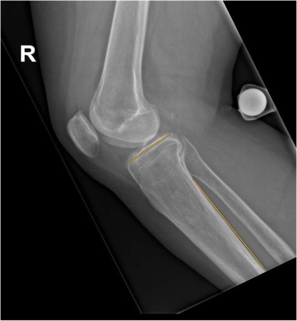
Background: The tibial slope plays an important role in knee surgery. However, standard radiographic measurement techniques have a low reproducibility and do not allow differentiation between medial and lateral articular surfaces. Despite availability of three-dimensional imaging, so far, no real 3D measurement technique was introduced and compared to radiographic measurement, which were the purposes of this study.
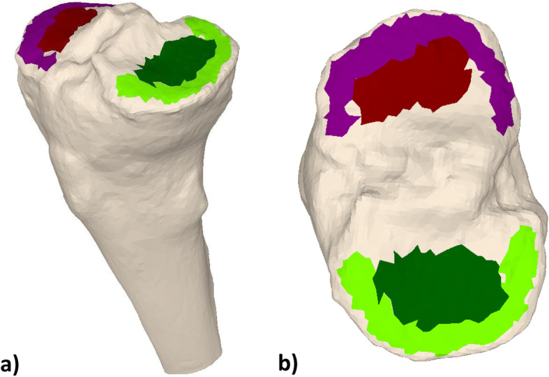
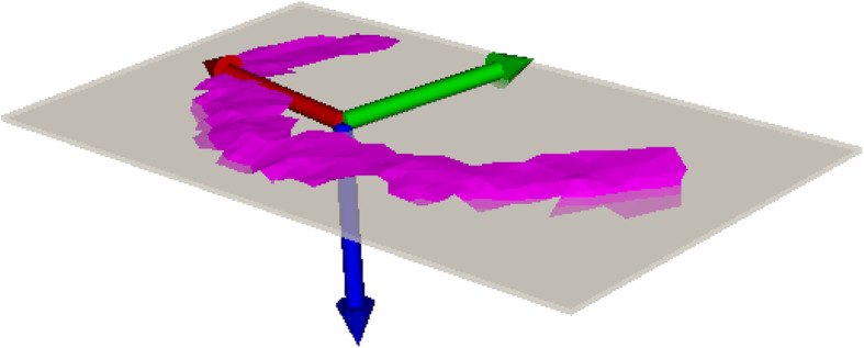
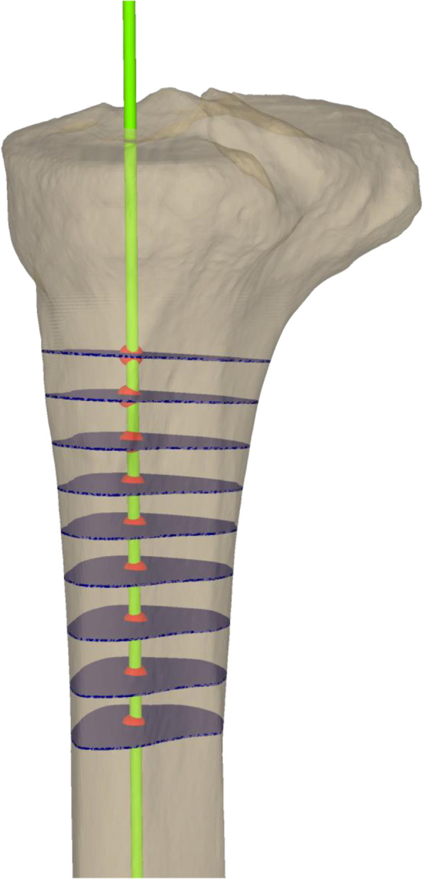
Methods: Computed tomography scans of 54 knees in 51 patients (41 males and 10 females) with a mean age of 46 years (range 22-67 years) were included. A novel 3D measurement technique was applied by two readers to measure the tibial slope of medial and lateral tibial plateau and rim. A statistical analysis was conducted to determine the intraclass correlation coefficient (ICC) for the new technique and compare it to a standard radiographic measurement.
Results: The mean 3D tibial slope for the medial plateau and rim was 7.4° and 7.6°, for the lateral plateau and rim 7.5° and 8.1°, respectively. The mean radiographic slope was 6.0°. Statistical analysis showed an ICC between both readers of 0.909, 0.987, 0.918, 0.893, for the 3D measurement of medial plateau, medial rim, lateral plateau and lateral rim, respectively, whereas the radiographic technique showed an ICC of 0.733.
Conclusions: The proposed novel measurement technique shows a high intraclass agreement and offers an applicable opportunity to assess the tibial slope three-dimensionally. Furthermore, the medial and lateral articular surfaces can be measured separately and one can differentiate the slope from the plateau and from the rim. As three-dimensional planning becomes successively more important, our measurement technique might deliver a useful supplement to the standard radiographic assessment in slope related knee surgery.
Level of evidence: Level III, diagnostic study.
Keywords: 3D measurement; Articular surface; Tibial slope.
Conflict of interest statement
The authors declare that they have no conflict of interest.
Figures
References
-
- Titze A. Variations in the slope of the proximal articular surface of the tibia. Z Orthop Ihre Grenzgeb. 1951;80(3):436–444. - PubMed
-
- Matsuda S, Miura H, Nagamine R, Urabe K, Ikenoue T, Okazaki K, et al. Posterior tibial slope in the normal and varus knee. Am J Knee Surg. 1999;12(3):165–168. - PubMed
-
- Walker PS, Garg A. Range of motion in total knee arthroplasty. A computer analysis. Clin Orthop Relat Res. 1991;(262):227–35. - PubMed
MeSH terms
Grants and funding
LinkOut - more resources
Full Text Sources
Research Materials
