. 2021 Jan 1:469:12-25.
doi: 10.1016/j.ydbio.2020.09.014. Epub 2020 Sep 25.

The actin polymerization factor Diaphanous and the actin severing protein Flightless I collaborate to regulate sarcomere size

Affiliations

The actin polymerization factor Diaphanous and the actin severing protein Flightless I collaborate to regulate sarcomere size

Su Deng et al. Dev Biol. .

Abstract

The sarcomere is the basic contractile unit of muscle, composed of repeated sets of actin thin filaments and myosin thick filaments. During muscle development, sarcomeres grow in size to accommodate the growth and function of muscle fibers. Failure in regulating sarcomere size results in muscle dysfunction; yet, it is unclear how the size and uniformity of sarcomeres are controlled. Here we show that the formin Diaphanous is critical for the growth and maintenance of sarcomere size: Dia sets sarcomere length and width through regulation of the number and length of the actin thin filaments in the Drosophila flight muscle. To regulate thin filament length and sarcomere size, Dia interacts with the Gelsolin superfamily member Flightless I (FliI). We suggest that these actin regulators, by controlling actin dynamics and turnover, generate uniformly sized sarcomeres tuned for the muscle contractions required for flight.

Keywords: Actin filaments; Actin polymerization; Actin severing; Diaphanous; Drosophila; Flight muscle; Flightless I; Formins; Gelsolin; Muscle maintenance; Sarcomere.


Conflict of interest statement

Declaration of competing interest None.

Figures
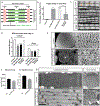
Figure 1:
Figure 1:. Knockdown of Diaphanous impairs flight ability and sarcomere size in IFMs
A. Schematic diagram of the structure of the sarcomere, noting Z disc, M line, thin and thick filaments. Length and width of the sarcomere are indicated. B. Flight assay: 25 flies per replicate, 3 replicates per genotype, n= 75 flies. The flight ability of individual flies was tested 3 days after eclosion. dia RNAi constructs were expressed in muscles with UAS-dicer2;;DMef2-Gal4. Reduced Dia activity resulted in impaired flight ability (p<0.0001). Rescue experiments were performed by expressing Dia::GFP in muscles along with dia RNAi (TRiP). The ability to fly was partially rescued by expressing Dia::GFP (p<0.0001). C. Myofibril and Sarcomere organization in flight muscle. Sarcomere length is indicated by the horizontal arrows, and sarcomere width is marked by the vertical arrows. Scale bar: 5μm; D. Quantification of sarcomere length and width: 5 flies per genotype, n = 5 average sarcomere length or width measurements per fly. In muscles with reduced Dia activity, both sarcomere length and width are significantly reduced (p<0.0001). Expressing Dia::GFP in the muscles restores flight capability, sarcomere length and width. E. Muscle cross sections indicating muscle size and myofibril organization. Scale bar: 10μm. F. Quantification of DLM3 cross-sectional areas: DLM3 from 5 flies per genotype, control n = 6 and dia-RNAi = 7 DLM3 muscles). In muscles with reduced Dia activity, muscle area is smaller than that of the controls, *p<0.05). G. Quantification of flight muscle myofibril area: 5 flies per genotype, 5 average myofibril area measurements per fly, n = 25 average myofibril area measurements per genotype. In muscles with reduced Dia activity, myofibril area is significantly smaller than that of the controls (****p<0.0001). H. Sarcomere morphology of newly eclosed adults using Transmission Electron Microscopy (TEM). Dia knockdown results in shorter and thinner sarcomeres. The numbers of thin and thick filaments are reduced. Scale bar: longitudinal sections: 600nm, cross sections: 200nm.
Figure 2:
Figure 2:. Knock down of Diaphanous impairs thin filament growth in IFMs.
A-B. Changes in IFM sarcomere size in wild-type adult flies. Wild-type flies were dissected at days 1 (newly eclosed), 3, and 7 after eclosion. The length and width of the individual sarcomeres was measured and recorded (5 flies per genotype, n = 5 average sarcomere length or width measurements/fly). Sarcomeres in newly eclosed flies are significantly shorter (p<0.0001) and thinner (p<0.0001) than the sarcomeres of flies 3 days after eclosion. There is no significant difference in sarcomere size between day 3 and day 7 flies. These data are consistent with continued growth of the sarcomeres in newly eclosed flies (day 1), followed by sarcomere maintenance (thereafter). Scale bar: 5μm C-D. Sarcomere phenotypes upon Dia knockdown in the muscle. Sarcomere size was measured on Day 1, 3 and 7 after eclosion (5 flies per genotype, n = 5 average sarcomere length or width measurements/fly). There is no significant difference in sarcomere size between each group. E. Diagram of experimental design. dia-RNAi was induced in adult muscle 3 days after eclosion. F. Viability of adult fly was examined after induction of dia-RNAi. No significant difference in viability was observed between control (n = 56 flies) and Dia knockdown (n = 52 flies) groups at the indicated time points. G. Quantification of Flight Performance following Dia knockdown. Flight performance was measured after 0, 4 and 11 days of induced dia-RNAi (3, 7, and 14 days after eclosion). Difference of flight performance was observed between control (Day 0; n = 140 flies, Day 4; n =51 flies, Day 7;n = 103 flies) and Dia knockdown (Day 0; n = 67 flies, Day 4; n = 54 flies, Day 7;n = 85 flies) groups following 4 (p<0.05) and 11 (p<0.0001) days of induction. H-J. Sarcomere morphology and size was examined after dia-RNAi induction. Average myofibril measurements were made at the indicated days of knockdown induction. Four days after induced Dia knockdown, sarcomere length and width are significantly reduced compare to control (5 flies per genotype, n = 5 average sarcomere length or width measurements/fly, ****p<0.0001). Scale bar: 2μm.
Figure 3:
Figure 3:. Knock down of Diaphanous results in changes in the localization of thin filament capping proteins.
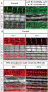
A. CapZ localization upon Dia knockdown in the IFMs. Flies were dissected 3 days after eclosion. 5 flies per genotype were dissected. 4 myofibrils/fly were imaged for analysis. Muscles are labeled with Phalloidin (white) and with an antibody against Cpa, a subunit of CapZ. B-C. Tmod localization in the IFMs upon dia knockdown. Flies were dissected 1 and 3 days after eclosion. 5 flies per genotype were dissected. 4 myofibrils/fly were imaged for analysis. Muscles are labeled with Phalloidin (white) and with an antibody against Tmod (red). Scale bar: 5 μm.
Figure 4:
Figure 4:. Diaphanous localization in IFM sarcomeres.
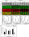
A. Dia localization in flies from day 1 (i), 3 (iii) and 7(v) after eclosion. Sarcomere thin filaments are labeled with Phalloidin (white). Z-discs are labeled with an antibody against α-actinin (red). Dia localization is visualized by staining with an antibody against Dia (green). Dia localizes mainly to the M-lines (arrow) 1 day after eclosion, and then shifts to the Z-discs as the sarcomeres cease growing (arrow head). The shift in Dia localization is verified by plotting the fluorescent signals in the boxed area: day 1 (ii), day 3 (iv) and day 7 (vi). B. Quantification of Dia localization in adult flies 1–7 days after eclosion. Fluorescence intensities of Dia were measured at the Z-disc, M-line, and at the area between the two regions from immunostained images. The relative percentage of Dia signal at the Z-disc and M-line was calculated and plotted (5 flies per genotype, 3 sarcomere signal measurements per fly, n = 15 sarcomere signal measurements, **** p<0.0001).
Figure 5:
Figure 5:. Dia and FliI genetically interact to regulate sarcomere size.
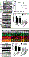
A. Sarcomere phenotype in muscles upon FliI knockdown. Flies were dissected 3 days after eclosion. Myofibrils are stained with Phalloidin (white), myofibrils without M-lines are marked with an arrow. Short sarcomeres are marked with an arrowhead and shown in the enlarged images. The bottom panel shows the sarcomere phenotype in flies that are rescued with FliI::HA. B. Survival assay. 25 embryos/genotype/day are selected for 3 days. Survival rates were calculated at the larval, pupal and adult stages. Knockdown of both Dia and FliI results in 100% lethality at the pupal stage. C. Sarcomere phenotypes in the pupae. 100 APF pupae were dissected. The morphology of myofibrils and sarcomeres are visualized with Phalloidin (white) Scale bar: 5 μm D. Quantification of sarcomere size. Knockdown of both FliI and Dia significantly reduces sarcomere length (p<0.01) and width (p<0.0001) compared to individually depleting either of the 2 proteins (5 flies per genotype, n = 5 average sarcomere length or width measurements/fly). E. Localization of FliI in an IFM. Control flies were dissected 1 to 7 days after eclosion. Muscles are labeled with Phalloidin (white) and with an antibody against α-actinin (red). The localization of FliI is visualized with a FliI antibody (green), which co-localizes with α-actinin at the Z-discs. Scale bars: 5 μm. F. Sarcomere phenotype in IFMs with FliI overexpression and Dia knockdown. Morphology of myofibrils and sarcomeres are visualized by labeling with Phalloidin (white). Scale bar: 5μm. G. Quantification of sarcomere size (5 flies per genotype, n = 5 average sarcomere length or width measurements/fly). Expressing both dia RNAi and FliI::HA in the muscles with two copies of DMef2-Gal4 significantly reduces sarcomere length (p<0.001) and width (p<0.1) in comparison to dia RNAi or FliI::HA alone.
Figure 6:
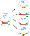
Figure 6:. Model: Dia and FliI collaborate to regulate sarcomere size and uniformity.
Under wild-type conditions, Dia and FliI act upon actin thin filaments during sarcomere growth to determine sarcomere size. Dia nucleates and polymerizes actin filaments, while FliI severs actin filaments. Together, Dia and FliI regulate the dynamics of actin thin filaments and maintain the balance between the F-actin and G-actin pools. Without Dia activity, actin filament elongation is impaired and the sarcomeres are shorter and thinner when compared to the control. Without FliI activity, long F-actin bundles form as a result of impaired actin-severing activity. Reduced actin severing also decreases the G-actin pool that limits actin polymerization by Diaphanous into the thin filament. As a result, the sarcomeres are shorter and thinner in comparison to the wild-type control. Knockdown of both FliI and Dia results in long F-actin bundles that form due to reduced actin severing activity and polymerization by residual Dia or other formins; however, the sarcomeres are even smaller than the single knock downs, due to both reduced G-actin pools and reduced actin elongation. When FliI is overexpressed in a background where Dia is knocked down, excessive actin severing and insufficient actin elongation results in sarcomeres that are smaller than just by manipulating either of the proteins individually.

Similar articles

Cited by

References

    1. Afshar K, Stuart B, Wasserman SA, 2000. Functional analysis of the Drosophila diaphanous FH protein in early embryonic development. Development 127, 1887–1897. - PubMed
    1. Arimura T, Takeya R, Ishikawa T, Yamano T, Matsuo A, Tatsumi T, Nomura T, Sumimoto H, Kimura A, 2013. Dilated cardiomyopathy-associated FHOD3 variant impairs the ability to induce activation of transcription factor serum response factor. Circ J 77, 2990–2996. - PubMed
    1. Babcock DT, Ganetzky B, 2014. An improved method for accurate and rapid measurement of flight performance in Drosophila. J Vis Exp, e51223. - PMC - PubMed
    1. Bai J, Hartwig JH, Perrimon N, 2007. SALS, a WH2-domain-containing protein, promotes sarcomeric actin filament elongation from pointed ends during Drosophila muscle growth. Dev Cell 13, 828–842. - PubMed
    1. Balakrishnan M, Yu SF, Chin SM, Soffar DB, Windner SE, Goode BL, Baylies MK, 2020. Cofilin Loss in Drosophila Muscles Contributes to Muscle Weakness through Defective Sarcomerogenesis during Muscle Growth. Cell Rep 32, 107893. - PMC - PubMed

Publication types

LinkOut - more resources