. 2021 Mar;160(4):1208-1223.e4.
doi: 10.1053/j.gastro.2020.09.030. Epub 2020 Sep 24.

Sympathetic Input to Multiple Cell Types in Mouse and Human Colon Produces Region-Specific Responses

Affiliations

Sympathetic Input to Multiple Cell Types in Mouse and Human Colon Produces Region-Specific Responses

Kristen M Smith-Edwards et al. Gastroenterology. 2021 Mar.

Abstract

Background & aims: The colon is innervated by intrinsic and extrinsic neurons that coordinate functions necessary for digestive health. Sympathetic input suppresses colon motility by acting on intrinsic myenteric neurons, but the extent of sympathetic-induced changes on large-scale network activity in myenteric circuits has not been determined. Compounding the complexity of sympathetic function, there is evidence that sympathetic transmitters can regulate activity in non-neuronal cells (such as enteric glia and innate immune cells).

Methods: We performed anatomical tracing, immunohistochemistry, optogenetic (GCaMP calcium imaging, channelrhodopsin), and colon motility studies in mice and single-cell RNA sequencing in human colon to investigate how sympathetic postganglionic neurons modulate colon function.

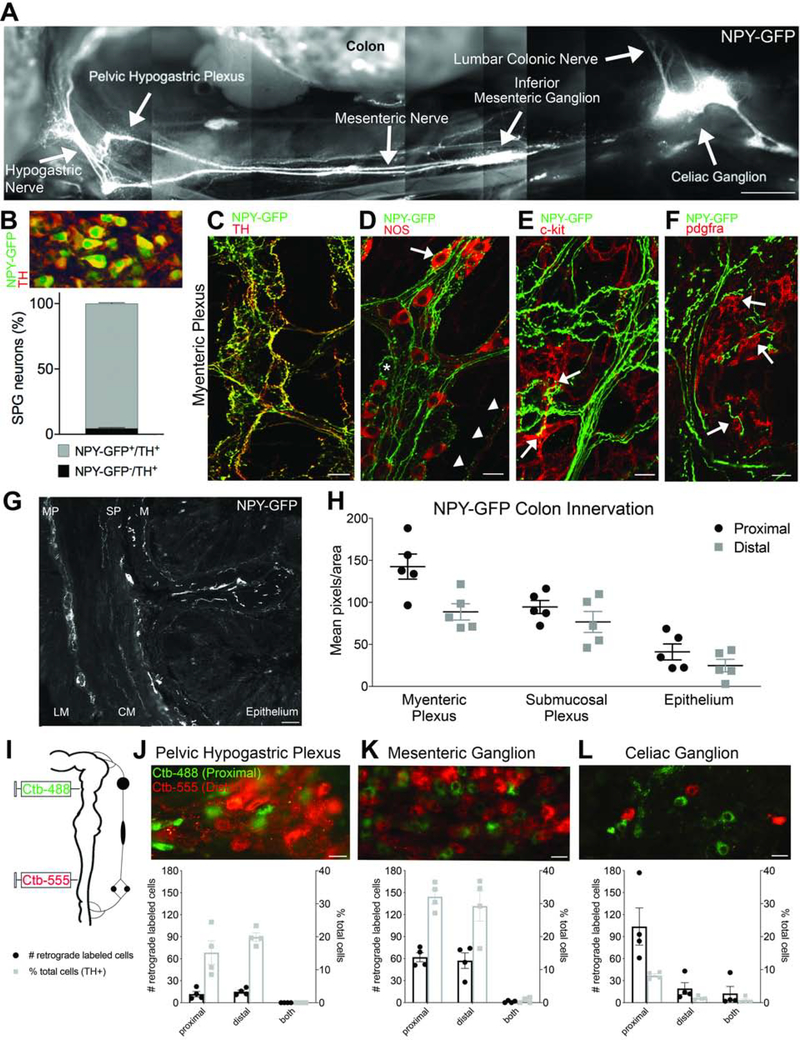
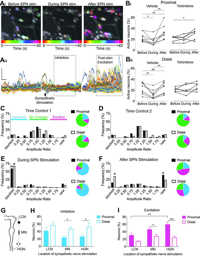
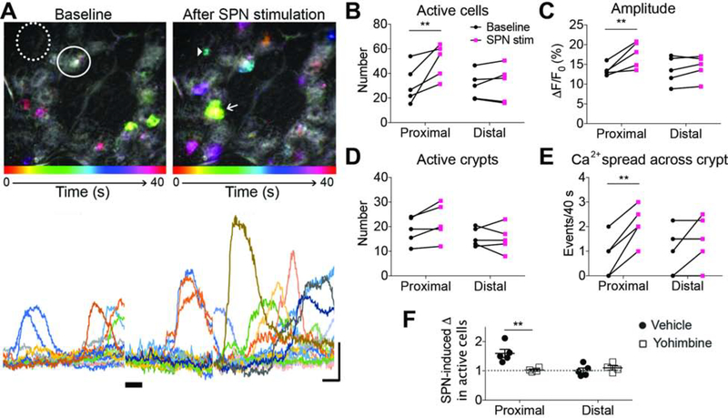
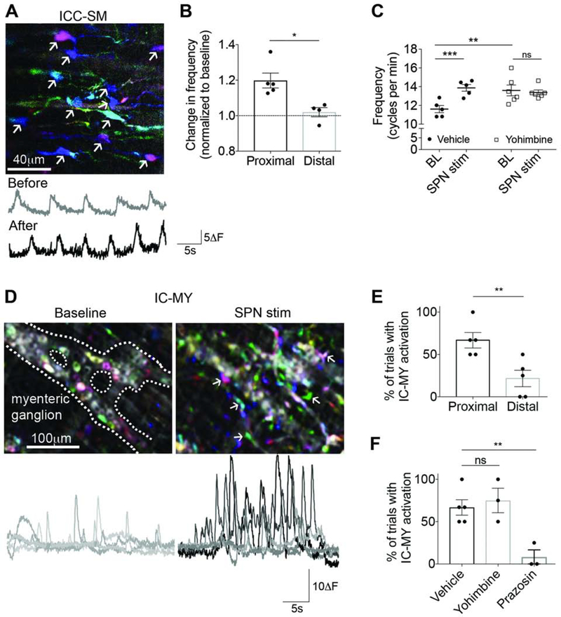
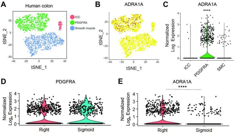
Results: Individual neurons in each sympathetic prevertebral ganglion innervated the proximal or distal colon, with processes closely opposed to multiple cell types. Calcium imaging in semi-intact mouse colon preparations revealed changes in spontaneous and evoked neural activity, as well as activation of non-neuronal cells, induced by sympathetic nerve stimulation. The overall pattern of response to sympathetic stimulation was unique to the proximal or distal colon. Region-specific changes in cellular activity correlated with motility patterns produced by electrical and optogenetic stimulation of sympathetic pathways. Pharmacology experiments (mouse) and RNA sequencing (human) indicated that appropriate receptors were expressed on different cell types to account for the responses to sympathetic stimulation. Regional differences in expression of α-1 adrenoceptors in human colon emphasize the translational relevance of our mouse findings.

Conclusions: Sympathetic neurons differentially regulate activity of neurons and non-neuronal cells in proximal and distal colon to promote distinct changes in motility patterns, likely reflecting the distinct roles played by these 2 regions.

Keywords: GCaMP; Gastrointestinal; Interstitial Cells of Cajal; Platelet-Derived Growth Factor Receptor–α.


Conflict of interest statement

All authors have no conflict of interest.

Figures

Figure 1.
Figure 1.. Individual sympathetic postganglionic neurons (SPNs) innervate proximal or distal colon.
(A) Montage shows innervation from sympathetic prevertebral ganglia to colon in NPY-GFP reporter mouse. (B) Quantification of sympathetic ganglion neurons that express NPY-GFP and/or TH (all NPY-GFP+ cells expressed TH). (C) Whole-mount immunolabeling reveals colocalization of TH and NPY-GFP+ fibers. (D) NPY-GFP+ fibers appear to innervate NOS+ (arrow) and NOS- myenteric neurons (asterisk); NPY-GFP+ varicosities are also closely opposed to NOS+ terminals within smooth muscle (arrowheads). (E-F) NPY-GFP+ fibers make close contacts to c-kit+ interstitial cells of Cajal (E) and PDGFRA+ cells (F) in the myenteric plexus. (G) Cross section of NPY-GFP mouse colon shows innervation to all layers; LM, longitudinal muscle, MP, myenteric plexus, CM, circular muscle, SP, submucosal plexus, M, mucosa, E, epithelium. (H) Comparison of NPY-GFP fiber density in proximal and distal colon. (I) Colon back-labeling experimental design. (J-L) Individual sympathetic postganglionic neurons in the pelvic hypogastric plexus (J), inferior mesenteric ganglion (K), and celiac ganglion (L) project to proximal (green) or distal (red) colon regions. Bar graphs show total number (left axis, black) and percentage (right axis, gray) of labeled cells. Scale bars, 1mm (A), 100μm (G), 20μm (C-F,J-L).
Figure 2.
Figure 2.. Electrical and optogenetic stimulation of SPNs produce local and pan-colonic motility changes.
(A) Maximum intensity projection of GCaMP fluorescence of SPNs. (B) Example traces of spontaneous activity in SPNs. (C) Electrical stimulation (20Hz) of LCN, MN, and HGN differentially activated neurons in sympathetic prevertebral ganglia; note that only MN stimulation was effective at producing responses (ΔF) in all three ganglia. (D) Amplitude of SPN responses increased as stimulation frequency increased. Inset, example GCaMP traces from an individual SPN to electrical stimulation of the MN at 1–20 Hz for 5 s (black bar); (E) Average tissue movement before, during, and after nerve stimulation at 1–20 Hz. Movement was significantly decreased during electrical stimulation (ES). (F) Experimental design to measure motility changes produced by optogenetic (blue laser) stimulation of SPNs using NPY-ChR2 mice. (G) Laser also significantly decreased local motility during stimulation. (H-J) Effects of ES on pan-colonic motility patterns. (K-M) Effects of laser on pan-colonic motility patterns. *p<0.05, **p<0.01, ***p<0.001, repeated measures (rm) two-way ANOVA, Tukey’s post-hoc correction for multiple comparisons (D,E,G); paired Student’s t-test (H-M). Scale bars, 20ΔF, 1s (B,D).
Figure 3.
Figure 3.. Sympathetic input differentially influences myenteric neuron activity in proximal and distal colon partially mediated via alpha-2 adrenergic receptors.
(A) Time-lapse color-coded images (Ai) and F/F traces (Aii) of GCaMP activity in myenteric neurons before, during, and after SPN stimulation (black arrow). Gray boxes highlight two phases of response (inhibition and post-stimulation excitation) to sympathetic activation. White arrows indicate neurons with increased spontaneous activity and arrowheads indicate neurons with “new” activity. (B) Percentage of myenteric neurons in proximal (Bi) and distal (Bii) colon with GCaMP activity before, during, and after SPN stimulation in the presence of vehicle (left) or yohimbine (right). (C-F) Histograms and corresponding pie charts showing the relative frequency of neurons in proximal (black) and distal (white) colon exhibiting changes in amplitude of GCaMP signals over time (C and D), during SPN stimulation (E), and after SPN stimulation (F); frequency of blocked neurons during stimulation was significantly higher compared to time-controls in both regions, but after stimulation, proximal colon had more “new” active neurons, whereas more neurons in the distal colon had sustained inhibition compared to time-controls. (G) Schematic illustrates nerves from SPGs to the colon. (H-I) Comparison of the frequency of neurons in the proximal and distal colon that are inhibited (H) or excited (I) after LCN, MN, and HGN stimulation. *p<0.05, **p<0.01, ***p<0.001; rm one-way ANOVA (B) and rm two-way ANOVA (H,I), Tukey’s post-hoc correction for multiple comparisons.
Figure 4.
Figure 4.. Sympathetic input has region-specific effects on ascending and descending myenteric neuron circuits.
(A) Diagram illustrating experimental setup. (B-C) Percentage of myenteric neurons in proximal (B) and distal (C) colon that responded to colon stimulation in isolated colon preparations (circles) and colon preparations with SPG intact (without external SPN activation; squares). (D-F) Percentage of neurons in proximal (D), middle (E), and distal (F) colon regions that responded to oral (i) or anal (ii) stimulation during and immediately following SPN stimulation. *p<0.05, **p<0.01; two-way ANOVA (B,C) and one-way ANOVA (D-F) with Tukey’s post hoc test for multiple comparisons.
Figure 5.
Figure 5.. Sympathetic input selectively increases GCaMP activity in proximal colon epithelium.
(A) Time-lapse color-coded images (top) and traces (bottom) of GCaMP activity in epithelial cells before (left) and after (right) SPN stimulation. Solid circle, active crypt; dotted circle, inactive crypt; arrowhead, active epithelial cell; arrow, “calcium spread” across >2 cells. (B-E) The number of active epithelial cells (B), amplitude of GCaMP signal (C), number of active crypts (D), and number of ”calcium spread” events were measured before and after SPN stimulation and compared between the proximal and distal colon. (F) Effects of yohimbine on SPN-induced changes in active epithelial cells in proximal and distal colon; dotted at y=1 represents no change in activity. **p<0.01; rm two-way ANOVA (B,C,E), two-way ANOVA (F) with Tukey’s post hoc test for multiple comparisons.
Figure 6.
Figure 6.. Sympathetic input influences interstitial cells in proximal colon.
(A) Time-lapse color-coded images (left) and traces (right) of GCaMP activity from interstitial cells of Cajal in the submucosal plexus (ICC-SM) (arrows) before and after SPN stimulation. (B) SPN-induced changes in ICC-SM frequency were significantly different in proximal and distal colon. (C) In proximal colon regions, SPN stimulation significantly increased ICC-SM frequency (filled circles, vehicle); yohimbine increased baseline frequency but blocked the response to SPN stimulation (open squares). (D) Time-lapse color-coded images (top) and traces (bottom) of GCaMP activity from interstitial cells in the myenteric plexus (IC-MY) (arrows) at baseline and with SPN stimulation. (E) Percentage of trials with SPN-induced activation of IC-MY in proximal colon was significantly greater than distal colon. (F) Yohimbine did not prevent IC-MY activation, but alpha-1 receptor antagonist, prazosin, significantly decreased IC-MY activation due to SPN stimulation. *p<0.05, **p<0.01, ***p<0.001; Student’s unpaired t-test (B,E), two-way (C) and one-way (F) ANOVA with Tukey’s post hoc test for multiple comparisons.
Figure 7.
Figure 7.. RNA sequencing of human interstitial cells provides molecular evidence that region-specific responses to sympathetic input exist in human colon.
(A) T-SNE plot showing 1684 SIP syncytium nuclei from human colon; ICC, PDGFRA+, and smooth muscle cells (SMC) formed distinct clusters, identified using canonical markers (Supplementary Figure S7). (B-C) T-SNE and violin plots showing the adrenoceptor, ADRA1A, was highly expressed in PDGFRA+ cells, but not in other groups. (D-E) ADRA1A expression was significantly higher in right colon compared to sigmoid colon, despite no difference in PDGFRA. ***p< 0.0001; Wilcoxon rank sum test with Bonferroni correction (C,E).

Similar articles

Cited by

References

    1. Schneider S, Wright CM, Heuckeroth RO. Unexpected Roles for the Second Brain: Enteric Nervous System as Master Regulator of Bowel Function. Annu Rev Physiol 2019;81:235–259. - PubMed
    1. Iino S, Ward SM, Sanders KM. Interstitial cells of Cajal are functionally innervated by excitatory motor neurones in the murine intestine. J Physiol 2004;556:521–30. - PMC - PubMed
    1. Baker SA, Drumm BT, Cobine CA, et al. Inhibitory Neural Regulation of the Ca (2+) Transients in Intramuscular Interstitial Cells of Cajal in the Small Intestine. Front Physiol 2018;9:328. - PMC - PubMed
    1. Baker SA, Drumm BT, Skowronek KE, et al. Excitatory Neuronal Responses of Ca(2+) Transients in Interstitial Cells of Cajal in the Small Intestine. eNeuro 2018;5. - PMC - PubMed
    1. Bellono NW, Bayrer JR, Leitch DB, et al. Enterochromaffin Cells Are Gut Chemosensors that Couple to Sensory Neural Pathways. Cell 2017;170:185–198 e16. - PMC - PubMed

Publication types

MeSH terms

LinkOut - more resources