. 2021 May;128(5):693-705.
doi: 10.1016/j.ophtha.2020.09.025. Epub 2020 Sep 24.

Quantitative Analysis of OCT for Neovascular Age-Related Macular Degeneration Using Deep Learning

Affiliations

Quantitative Analysis of OCT for Neovascular Age-Related Macular Degeneration Using Deep Learning

Gabriella Moraes et al. Ophthalmology. 2021 May.

Abstract

Purpose: To apply a deep learning algorithm for automated, objective, and comprehensive quantification of OCT scans to a large real-world dataset of eyes with neovascular age-related macular degeneration (AMD) and make the raw segmentation output data openly available for further research.

Design: Retrospective analysis of OCT images from the Moorfields Eye Hospital AMD Database.

Participants: A total of 2473 first-treated eyes and 493 second-treated eyes that commenced therapy for neovascular AMD between June 2012 and June 2017.

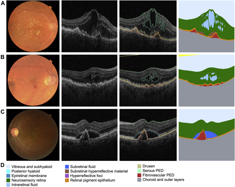
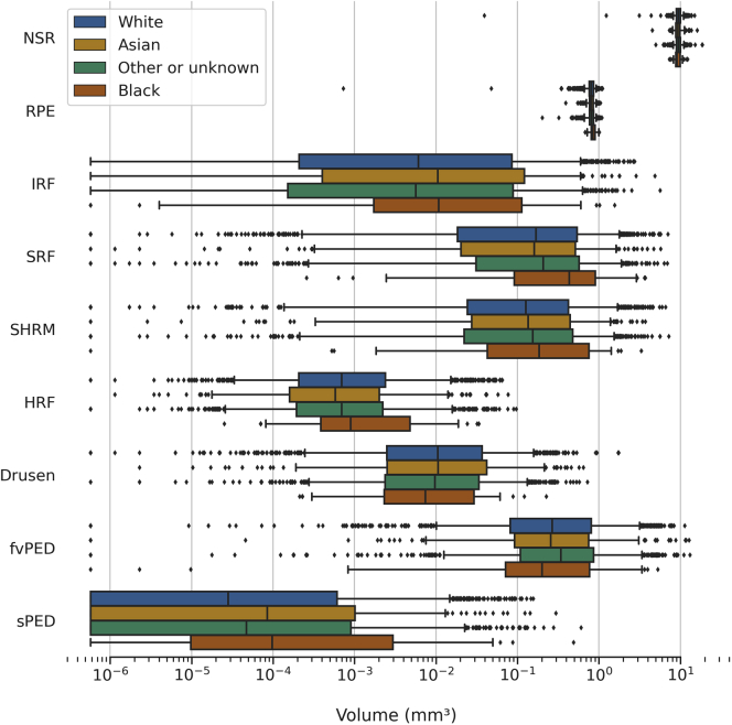
Methods: A deep learning algorithm was used to segment all baseline OCT scans. Volumes were calculated for segmented features such as neurosensory retina (NSR), drusen, intraretinal fluid (IRF), subretinal fluid (SRF), subretinal hyperreflective material (SHRM), retinal pigment epithelium (RPE), hyperreflective foci (HRF), fibrovascular pigment epithelium detachment (fvPED), and serous PED (sPED). Analyses included comparisons between first- and second-treated eyes by visual acuity (VA) and race/ethnicity and correlations between volumes.

Main outcome measures: Volumes of segmented features (mm3) and central subfield thickness (CST) (μm).

Results: In first-treated eyes, the majority had both IRF and SRF (54.7%). First-treated eyes had greater volumes for all segmented tissues, with the exception of drusen, which was greater in second-treated eyes. In first-treated eyes, older age was associated with lower volumes for RPE, SRF, NSR, and sPED; in second-treated eyes, older age was associated with lower volumes of NSR, RPE, sPED, fvPED, and SRF. Eyes from Black individuals had higher SRF, RPE, and serous PED volumes compared with other ethnic groups. Greater volumes of the majority of features were associated with worse VA.

Conclusions: We report the results of large-scale automated quantification of a novel range of baseline features in neovascular AMD. Major differences between first- and second-treated eyes, with increasing age, and between ethnicities are highlighted. In the coming years, enhanced, automated OCT segmentation may assist personalization of real-world care and the detection of novel structure-function correlations. These data will be made publicly available for replication and future investigation by the AMD research community.

Keywords: OCT; age-related macular degeneration; automated; deep learning; neovascular.


Figures

Figure 1
Figure 1
Fundus photograph, OCT scan, and corresponding segmentation map for 3 examples. A, Macular neovascularization (MNV) in a typical case of neovascular age-related macular degeneration (AMD): an 81-year-old White woman presenting with visual acuity (VA) of 63 Early Treatment Diabetic Retinopathy Study (ETDRS) letters. B, Type 3 MNV example: an 83-year-old woman of other/unknown ethnicity/race presenting VA of 70 ETDRS letters and OCT presenting intraretinal fluid (IRF) only. C, Polyp-like example in young patient: a 58-year-old Asian woman presenting VA of 59 ETDRS letters and OCT showing subretinal fluid (SRF) and suspicious polyp-like lesion. D, Color key for 13 anatomic features segmented by the segmentation network. PED = pigment epithelium detachment.
Figure 2
Figure 2
Distribution of segmented features volumes stratified by first- and second-treated eyes. The boxes show the median and interquartile range. Whiskers extend to the 5th and 95th percentiles, and beyond this outliers are shown individually. The volume (mm3) is distributed across a logarithmic scale; log(zero) is undefined, so zero values were set to the smallest positive value (5.8e-7). fvPED = fibrovascular pigment epithelium detachment; HRF = hyperreflective foci; IRF = intraretinal fluid; NSR = neurosensory retina; RPE = retinal pigment epithelium; SHRM = subretinal hyperreflective material; SRF = subretinal fluid; sPED = serous pigment epithelium detachment.
Figure 3
Figure 3
Spearman’s correlation between segmented feature volumes and central subfield thickness (CST) for (A) first- and (B) second-treated eyes. Tiles display the coefficient rs. The upper right half blanks out tiles that have a P > 0.05; values are symmetrical otherwise. fvPED = fibrovascular pigment epithelium detachment; HRF = hyperreflective foci; IRF = intraretinal fluid; NSR = neurosensory retina; RPE = retinal pigment epithelium; SHRM = subretinal hyperreflective material; sPED = serous pigment epithelium detachment; SRF = subretinal fluid.
Figure 4
Figure 4
Distribution of first-treated eye segmented feature volumes stratified by baseline visual acuity (VA) subgroups. The VA is stratified into Early Treatment Diabetic Retinopathy Study (ETDRS) letters of 0–35, 36–52, 53–69, and 70 or greater (Table S4, available at www.aaojournal.org). The boxes show the median and interquartile range. Whiskers extend to the 5th and 95th percentiles, and beyond this outliers are shown individually. The volume is distributed across a logarithmic scale; log(zero) is undefined, so zero values were set to the smallest positive value (5.8e-7). fvPED = fibrovascular pigment epithelium detachment; HRF = hyperreflective foci; IRF = intraretinal fluid; NSR = neurosensory retina; RPE = retinal pigment epithelium; SHRM = subretinal hyperreflective material; sPED = serous pigment epithelium detachment; SRF = subretinal fluid.
Figure 5
Figure 5
Distribution of segmented feature volumes in the first-treated eye at baseline stratified by age groups 50–59, 60–69, 70–79, and ≥80 years (Table S4, available at www.aaojournal.org) across a logarithmic scale; log(zero) is undefined, so zero values were set to the smallest positive value (5.8e-7). The boxes show the median and interquartile range. Whiskers extend to the 5th and 95th percentiles, and beyond this outliers are shown individually. fvPED = fibrovascular pigment epithelium detachment; HRF = hyperreflective foci; IRF = intraretinal fluid; NSR = neurosensory retina; RPE = retinal pigment epithelium; SHRM = subretinal hyperreflective material; sPED = serous pigment epithelium detachment; SRF = subretinal fluid.
Figure 6
Figure 6
Distribution of segmented feature volumes in the first-treated eyes at baseline stratified by ethnicities: White, Asian, other or unknown, and Black (Table S4, available at www.aaojournal.org). The boxes show the median and interquartile range. Whiskers extend to the 5th and 95th percentiles, and beyond this outliers are shown individually. The volume is distributed across a logarithmic scale; log(zero) is undefined, so zero values were set to the smallest positive value (5.8e-7). fvPED = fibrovascular pigment epithelium detachment; HRF = hyperreflective foci; IRF = intraretinal fluid; NSR = neurosensory retina; RPE = retinal pigment epithelium; SHRM = subretinal hyperreflective material; sPED = serous pigment epithelium detachment; SRF = subretinal fluid.

References

    1. Lai T.-T., Hsieh Y.-T., Yang C.-M. Biomarkers of optical coherence tomography in evaluating the treatment outcomes of neovascular age-related macular degeneration: a real-world study. Sci Rep. 2019;9:529. - PMC - PubMed
    1. Schmidt-Erfurth U., Waldstein S.M. A paradigm shift in imaging biomarkers in neovascular age-related macular degeneration. Prog Retin Eye Res. 2016;50:1–24. - PubMed
    1. Waldstein S.M., Philip A.-M., Leitner R. Correlation of 3-dimensionally quantified intraretinal and subretinal fluid with visual acuity in neovascular age-related macular degeneration. JAMA Ophthalmol. 2016;134:182–190. - PubMed
    1. Fung A.E., Lalwani G.A., Rosenfeld P.J. An optical coherence tomography-guided, variable dosing regimen with intravitreal ranibizumab (Lucentis) for neovascular age-related macular degeneration. Am J Ophthalmol. 2007;143:566–583. - PubMed
    1. Lalwani G.A., Rosenfeld P.J., Fung A.E. A variable-dosing regimen with intravitreal ranibizumab for neovascular age-related macular degeneration: year 2 of the PrONTO Study. Am J Ophthalmol. 2009;148:43–58.e1. - PubMed

Publication types