Review
. 2021 Feb;78(3):358-370.
doi: 10.1111/his.14264. Epub 2020 Dec 1.

Hunting coronavirus by transmission electron microscopy - a guide to SARS-CoV-2-associated ultrastructural pathology in COVID-19 tissues

Affiliations
Review

Hunting coronavirus by transmission electron microscopy - a guide to SARS-CoV-2-associated ultrastructural pathology in COVID-19 tissues

Helmut Hopfer et al. Histopathology. 2021 Feb.

Abstract

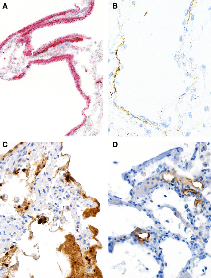
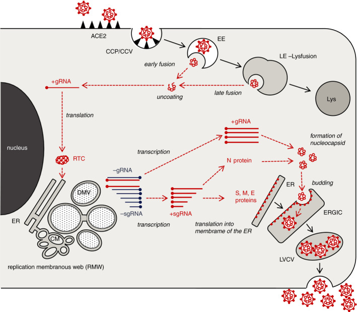
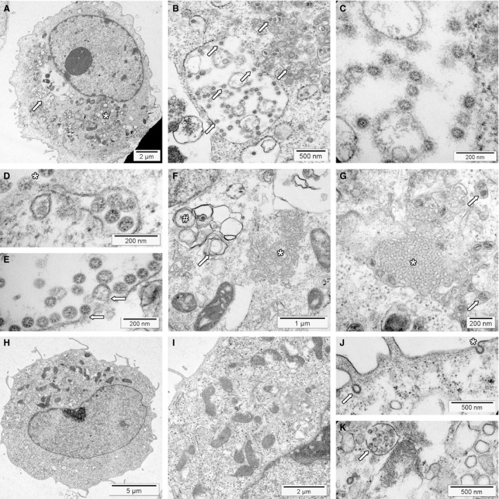
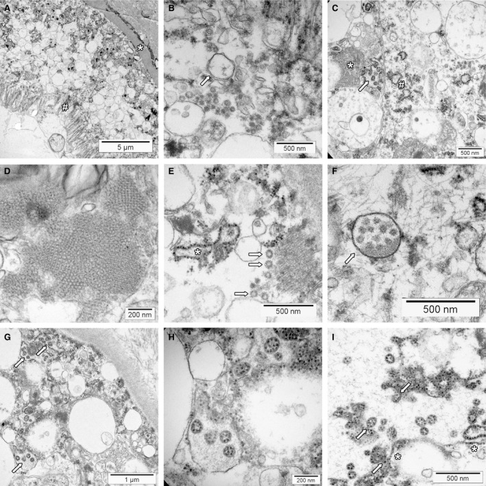
Transmission electron microscopy has become a valuable tool to investigate tissues of COVID-19 patients because it allows visualisation of SARS-CoV-2, but the 'virus-like particles' described in several organs have been highly contested. Because most electron microscopists in pathology are not accustomed to analysing viral particles and subcellular structures, our review aims to discuss the ultrastructural changes associated with SARS-CoV-2 infection and COVID-19 with respect to pathology, virology and electron microscopy. Using micrographs from infected cell cultures and autopsy tissues, we show how coronavirus replication affects ultrastructure and put the morphological findings in the context of viral replication, which induces extensive remodelling of the intracellular membrane systems. Virions assemble by budding into the endoplasmic reticulum-Golgi intermediate complex and are characterised by electron-dense dots of cross-sections of the nucleocapsid inside the viral particles. Physiological mimickers such as multivesicular bodies or coated vesicles serve as perfect decoys. Compared to other in-situ techniques, transmission electron microscopy is the only method to visualise assembled virions in tissues, and will be required to prove SARS-CoV-2 replication outside the respiratory tract. In practice, documenting in tissues the characteristic features seen in infected cell cultures seems to be much more difficult than anticipated. In our view, the hunt for coronavirus by transmission electron microscopy is still on.

Keywords: coronavirus; electron microscopy; ultrastructure; virus replication.


Conflict of interest statement

The authors declare no conflicts of interest.

Figures

Figure 1
Figure 1
In‐situ detection of severe acute respiratory syndrome (SARS)‐CoV‐2 RNA and proteins. In‐situ imaging of SARS‐CoV‐2 RNA and proteins reflects the disproportionate production of various virus RNA and antigen compounds. A, In‐situ hybridisation (ISH) applying the 845701 RNAscope probe – V‐nCoV2019‐S‐sense duplexed with the 859151 RNAscope probe – V‐nCoV2019‐orf1ab‐sense from Advanced Cell Diagnostics (Hayward, CA, USA) yields abundant SARS‐CoV‐2 RNA within the hyaline membranes of an affected lung. This labelling corresponds to viral RNA, but not to complete virions. B, ISH using the 845701 RNAscope probe – V‐nCoV2019‐S‐sense highlights SARS‐CoV‐2 RNA within alveolar walls, most probably attributable to viral RNA within endothelial cells. C, Immunohistochemistry (IHC) for SARS‐CoV‐2 nucleocapsid antigens with the polyclonal rabbit anti‐nucleocapsid antibody from SinoBiological (Wayne, PA, USA) labels excessive amounts of viral protein present within the hyaline membranes of an affected lung, but does not correspond to intact virions. D, IHC staining for SARS‐CoV‐2 S protein with the clone 007 rabbit anti‐spike antibody from SinoBiological is confined to alveolar vessels. C and D were performed on autopsy cases from our published cohort by Mattia Bugatti in the laboratory of Fabio Facchetti.
Figure 2
Figure 2
Cartoon of SARS‐CoV‐2 replication in cells. Severe acute respiratory syndrome (SARS)‐CoV‐2 infection and replication can be arbitrarily divided into three phases. (1) After docking to its receptor, the virus is internalised. Proteolytic cleavage of the S protein results in the fusion of the viral envelope with the endosomal membrane, followed by release and uncoating of the viral RNA (upper right). (2) The genomic viral RNA (+gRNA) is translated by ribosomes producing the replication–transcription complex. This initiates an extensive remodelling of intracellular membranes forming the replication membranous web where genomic and subgenomic viral RNA (+sgRNA) are generated (left). (3) The +sgRNA coding for the structural envelope proteins of the virus is directly translated into the membranes of the ER. The nucleocapsid protein assembles with the +gRNA to form the nucleocapsid. Its invagination into the ERGIC forms new virions residing in vesicles, which are subsequently released (lower right). Viral structures are shown in red, cell components in black. For clarity, ribosomes are not depicted. CCP, clathrin‐coated pit; CCV, clathrin‐coated vesicle; EE, early endosome; LE, late endosome; Lys, lysosome; RTC, replication–transcription complex; ER, endoplasmic reticulum; DMV, double membrane vesicle; CM, convoluted membranes; ERGIC, endoplasmic reticulum Golgi intermediate complex.
Figure 3
Figure 3
Transmission electron microscopy of severe acute respiratory syndrome (SARS)‐CoV‐2 infected and non‐infected Vero cells. Cell blocks were prepared from SARS‐CoV‐2 infected and non‐infected Vero cells (see Supporting information for details), processed for transmission electron microscopy according to standard procedures and investigated using an FEI Morgagni 268D transmission electron microscope (TEM). Unmarked and uncropped high‐resolution images of all panels are provided in the Supporting information. A, SARS‐CoV‐2‐infected Vero cell at low power. The replication organelle is visible below the nucleus (*). The cell contains a large virion‐containing vacuole (LVCV; arrow) shown at higher magnification in B. B, At higher magnification, a LVCV as well as smaller vesicles contain multiple assembled virions (some marked by arrows). C, High magnification depicts SARS‐CoV‐2 virions inside the LVCV. Within virions, the nucleocapsid is visible as small electron‐dense dots. Depending on the cross‐sectional plane, the centre can be electron lucent. The ‘corona’ formed by the S protein is not visible using standard staining protocols. D, High magnification of a vesicle containing virions docked to the cell surface (*) for virus release; vacuole just under the plasma membrane contains virions to be exocytosed. E, High magnification of LVCV showing virus budding (arrows) and assembled virions. F, Area with convoluted membranes (*) next to mitochondria and double membrane vesicles (#) with signs of deterioration (myelin figure, arrow). Nucleus is shown in the upper left corner under the figure label (F). G, Area showing a cubic membrane structures with membranes arranged in an ordered fashion (*). The surrounding cytoplasm contains several vesicles containing virions (arrows). H, Non‐infected Vero cell. The cytoplasm contains mitochondria, rough endoplasmic reticulum and few vesicles shown at higher magnification in panel I. I, Higher magnification of a non‐infected Vero cell. The intracellular membranes are not prominent. J, Surface of a non‐infected Vero cell showing a coated pit at the cell surface (*) and a coated vesicle (arrow). K, Deteriorated non‐infected cell with a multivesicular body (MVB; arrow) with intraluminal vesicles inside, mimicking an LVCV. Note that the intraluminal vesicles do not show the dots of the nucleocapsid. The structure next to the MVB is a lysosome.
Figure 4
Figure 4
Transmission electron microscopy of COVID‐19 autopsy tissue. Membrane alterations and mimickers of severe acute respiratory syndrome (SARS)‐CoV‐2. Tissues were collected during autopsies and fixed in 4% formalin for at least 72 h. Small tissue blocks were cut and transferred to 3% glutaraldehyde. Processing for transmission electron microscopy was performed according to standard procedures. The tissues were investigated with FEI Morgagni 268D transmission electron microscopy (TEM). Unmarked and uncropped high‐resolution images of all panels are provided in the Supporting information. A, Overview of kidney proximal tubular epithelial cells on top of basement membrane (*). The interstitium is seen in the upper right corner, the brush border at the apical surface (#). The cytoplasm contains multiple membrane‐limited vesicles. B, Higher magnification reveals that some of the vesicles have double membranes (arrows) suggesting the presence of double membrane vesicles. C, This area of a kidney tubular epithelial cell shows convoluted membranes (*) and dilated rough endoplasmic reticulum (#). Some of the larger vacuoles contain vesicles with prominent electron‐dense dots that have the same size as the ribosomes of the endoplasmic reticulum (arrow, ‘outside‐in’ ribosomes). D, Cubic membrane structures within a pneumocyte of the lung. E, Coated vesicles (arrows) are seen in the basal part of this kidney proximal tubular epithelial cell. The tubular basement membrane is sectioned in the upper right corner. The rough endoplasmic reticulum (*) is dilated. F, A multivesicular body within a podocyte of a glomerulus (arrow). Note that the intraluminal vesicles do not show electron‐dense dots. G, Vesicles with ‘outside‐in’ ribosomes (arrows) mimicking SARS‐CoV‐2 virions in a proximal tubule of the kidney. Some of the surrounding vesicles show ribosomes at the outside, which have the same size as the electron‐dense dots on the inside. The tubular basement membrane and collagen fibrils of the interstitium are visible in the upper right corner. H, Higher magnification of vesicles with ‘outside‐in’ ribosomes. The surrounding membrane has no ribosomes attached. I, Part of a larger vesicle with some ribosomes attached on the cytoplasmic side of the membrane (*). The vesicles with ‘outside‐in’ ribosomes seem to be generated by invagination of the membrane (arrows).

References

    1. Li Q, Guan X, Wu P et al. Early transmission dynamics in Wuhan, China, of novel coronavirus‐infected pneumonia. N. Engl. J. Med. 2020; 382; 1199–1207. - PMC - PubMed
    1. Huang C, Wang Y, Li X et al. Clinical features of patients infected with 2019 novel coronavirus in Wuhan. China. Lancet 2020; 395; 497–506. - PMC - PubMed
    1. Battegay M, Kuehl R, Tschudin‐Sutter S et al. 2019‐novel Coronavirus (2019‐nCoV): estimating the case fatality rate – a word of caution. Swiss Med. Wkly 2020; 150; w20203. - PubMed
    1. Leuzinger K, Roloff T, Gosert R et al. Epidemiology of SARS‐CoV‐2 emergence amidst community‐acquired respiratory viruses. J. Infect. Dis. 2020; 222; 1270–1279. - PMC - PubMed
    1. Menter T, Haslbauer JD, Nienhold R et al. Postmortem examination of COVID‐19 patients reveals diffuse alveolar damage with severe capillary congestion and variegated findings in lungs and other organs suggesting vascular dysfunction. Histopathology 2020; 77; 198–209. - PMC - PubMed