

Scanning electron microscopy of the surface epithelium of the bovine endometrium
- PMID: 32981737
- PMCID: PMC8009268
- DOI: 10.3168/jds.2020-18852
Scanning electron microscopy of the surface epithelium of the bovine endometrium
Abstract
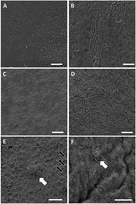
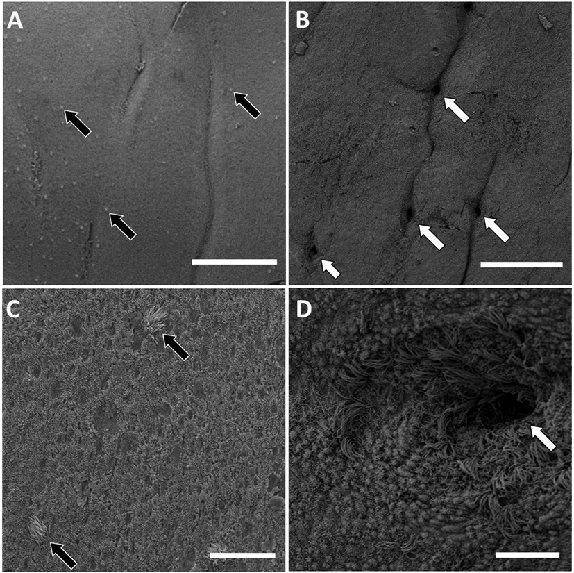
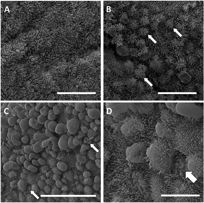
The surface epithelium of the bovine endometrium comprises at least 2 cell types (ciliated cells and secretory cells with microvilli), but their distribution and morphological changes over the estrous cycle are poorly understood. The objective was to quantify the number of ciliated cells and assess morphological changes in secretory cells on the uterine surface epithelium during the estrous cycle. Caruncular endometrium (CAR) and intercaruncular endometrium (ICAR) samples were collected from the uterine body, the horn ipsilateral to the corpus luteum or dominant follicle (H-CL/DF), and the horn contralateral to the corpus luteum or dominant follicle (H-NCL/NDF) from heifers following slaughter on d 0 (estrus; n = 5) or d 14 (mid-luteal phase; n = 5) of the estrous cycle. Samples were prepared for scanning electron microscopy at 1,000× magnification. Four to 10 fields (256 × 225 µm) for each sample were examined (n = 567 images). The number of ciliated cells was counted and the surface was scored for the morphology of the secretory cells (0 = absence of microvilli on surface; 3 = 100% of surface covered with microvilli). Ciliated cells were present in both the CAR and ICAR regions. The number of ciliated cells per field increased from d 0 to 14 in CAR and decreased from d 0 to14 in ICAR. The scanning electron microscopy revealed a general lack of uniformity in the lawn of microvilli on the surface of the endometrium. Based on the scores, approximately 25% of the fields had a surface that was <50% covered by microvilli. Depletion of microvilli may be explained by a normal process where apical protrusions are formed and either regress back into the cell surface or break to release their contents into the uterine lumen. These studies support the hypothesis that the surface of the luminal epithelium changes during the estrous cycle through a process that involves remodeling of the apical surface. The morphology of the apical surface may have a key role in governing pregnancy establishment.
Keywords: cilia; endometrium; microvilli; scanning electron microscopy.
Copyright © 2020 American Dairy Science Association. Published by Elsevier Inc. All rights reserved.
Figures
Similar articles
-
Differential gene expression profiling of endometrium during the mid-luteal phase of the estrous cycle between a repeat breeder (RB) and non-RB cows.Reprod Biol Endocrinol. 2017 Mar 23;15(1):20. doi: 10.1186/s12958-017-0237-6. Reprod Biol Endocrinol. 2017. PMID: 28335821 Free PMC article.
-
Endometrial effects of levonorgestrel and estradiol: A Scanning electron microscopic study of the luminal epithelium.Contraception. 1980 Jul;22(1):71-83. doi: 10.1016/0010-7824(80)90119-5. Contraception. 1980. PMID: 6774851
-
Cyclic changes in the surface structure of the cervix from the ewe as revealed by scanning electron microscopy.Tissue Cell. 1979;11(2):359-70. doi: 10.1016/0040-8166(79)90049-1. Tissue Cell. 1979. PMID: 572999
-
The effects of steroids on the fine structure of the endometrium.Baillieres Clin Obstet Gynaecol. 1989 Jun;3(2):227-48. doi: 10.1016/s0950-3552(89)80020-3. Baillieres Clin Obstet Gynaecol. 1989. PMID: 2692919 Review.
-
Microarchitecture of the human endometrium by scanning electron microscopy: menstrual desquamation and remodeling.Ann N Y Acad Sci. 1991;622:28-46. doi: 10.1111/j.1749-6632.1991.tb37848.x. Ann N Y Acad Sci. 1991. PMID: 2064187 Review.
Cited by
-
Uterine histomorphological and immunohistochemical investigation during the follicular phase of estrous cycle in Saidi sheep.BMC Vet Res. 2025 Jan 13;21(1):16. doi: 10.1186/s12917-024-04456-3. BMC Vet Res. 2025. PMID: 39806411 Free PMC article.
-
Establishment of the uterine microbiome following artificial insemination in virgin heifers.Front Microbiol. 2024 Jun 5;15:1385505. doi: 10.3389/fmicb.2024.1385505. eCollection 2024. Front Microbiol. 2024. PMID: 38903779 Free PMC article.
-
Transcriptome Analysis Reveals the Response Mechanism of Frl-Mediated Resistance to Fusarium oxysporum f. sp. radicis-lycopersici (FORL) Infection in Tomato.Int J Mol Sci. 2022 Jun 25;23(13):7078. doi: 10.3390/ijms23137078. Int J Mol Sci. 2022. PMID: 35806084 Free PMC article.
-
The impact of primary ciliary dyskinesia on female and male fertility: a narrative review.Hum Reprod Update. 2023 May 2;29(3):347-367. doi: 10.1093/humupd/dmad003. Hum Reprod Update. 2023. PMID: 36721921 Free PMC article. Review.
-
Extensive rewiring of the gene regulatory interactions between in vitro-produced conceptuses and endometrium during attachment.PNAS Nexus. 2023 Sep 2;2(9):pgad284. doi: 10.1093/pnasnexus/pgad284. eCollection 2023 Sep. PNAS Nexus. 2023. PMID: 37711857 Free PMC article.
References
-
- Almeida AP, Ayalon N, and Bartoov B. 1986. Bovine endometrial epithelium ultrastructure 6 and 7 days post-breeding. Animal Reproduction Science 10:293–300. doi:10.1016/0378-4320(86)90004-7. - DOI
MeSH terms
Grants and funding
LinkOut - more resources
Full Text Sources
Miscellaneous
