. 2020 Sep 14:13:2279-2287.
doi: 10.2147/JPR.S264255. eCollection 2020.

Ultrasound-Guided Unilateral Transversus Abdominis Plane Combined with Rectus Sheath Block versus Subarachnoid Anesthesia in Patients Undergoing Peritoneal Dialysis Catheter Surgery: A Randomized Prospective Controlled Trial

Affiliations

Ultrasound-Guided Unilateral Transversus Abdominis Plane Combined with Rectus Sheath Block versus Subarachnoid Anesthesia in Patients Undergoing Peritoneal Dialysis Catheter Surgery: A Randomized Prospective Controlled Trial

Ji Li et al. J Pain Res. .

Abstract

Background: Peritoneal dialysis catheter placement can be performed under general anesthesia, local anesthesia or subarachnoid anesthesia (SA). Recently, studies have reported the successful placement of peritoneal dialysis catheters using a transversus abdominis plane (TAP) block and rectus sheath (RS) block. This study compared the TAP + RS block with SA for patients undergoing peritoneal dialysis catheter placement.

Methods: Sixty patients were randomly divided into two groups, with 30 receiving unilateral ultrasound-guided TAP + RS block anesthesia and 30 receiving SA. The demographic characteristics, anesthesia efficacy, indicators related to anesthesia or operation, hemodynamic index, postoperative pain numeric rating score (NRS), postoperative recovery indicators, complications related to anesthesia or surgery, and dosage of sedative or analgesic medication were analyzed.

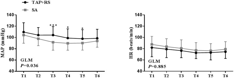
Results: Anesthesia operation time was significantly shorter in the TAP + RS block group than in the SA group (P<0.001), while there was no significant difference in success rates (TAP + RS 93.33% [95% confidence interval, 95% CI, 83.9-102.8%] vs SA 100.00% [95% CI, 100-100%], P=0.472). Two patients in the TAP + RS group needed extra analgesia, although the dermatome pinprick sensation test gave negative results for all patients. Patients who received the TAP + RS block expressed significantly less pain on movement or at rest at 4 h and 8 h postoperative. Fewer patients needed rescue analgesia with tramadol in the postoperative period in the TAP + RS block group than in the SA group (P<0.05). The intraoperative MAP was more stable (P<0.05) in the TAP + RS group compared to the SA group.

Conclusion: The TAP + RS block is a safe, effective method for use as the principal anesthesia technique in PD catheter placement. Compared to SA, it has the advantages of less influence on hemodynamics and a better postoperative analgesic effect.

Keywords: TAP block; local anesthesia; peritoneal dialysis catheter placement; rectus sheath block; subarachnoid anesthesia.


Conflict of interest statement

The authors report no conflicts of interest for this work.

Figures

Figure 1
Figure 1
CONSORT flowchart.
Figure 2
Figure 2
PD catheter surgery was completed. The yellow dotted line indicates the intraperitoneal indwelled tube. The red dotted line shows the peritoneal dialysis tube passing through the subcutaneous tunnel. The yellow arrow indicates the surgical incision.
Figure 3
Figure 3
(A) When placing the ultrasound linear probe at the level of anterior axillary line between the costal margin and the iliac crest, MOE, MOI and MTA are displayed legibly. The plane indicated by the red arrow is the fascia of the transverse abdominis, which is a potential space and the target plane of our injection. (B) The block is performed using in-plane techniques. After ropivacaine solution was injected into the fascia of the transverse abdominis, the fluid diffused into the fascial lumen, and the MTA was separated from the MOI and was pressed deep. The fluid dark area circled by the red dotted line indicates the diffusion of ropivacaine solution in the TAP.
Figure 4
Figure 4
When placing the ultrasound linear probe at the skin surface of the rectus abdominis above the umbilicus level, the RA could be displayed legibly and the RS is shown as a highlighted layer of fascia surrounding the RA. The posterior RS is a narrow space, representing the target injection site. After ropivacaine solution was injected into the posterior RS, the fluid diffused into a spindle shape within the fascial cavity. The fluid dark area circled by the red dotted line indicates the diffusion of ropivacaine solution into the RS. Abbreviations: RA, rectus abdominis; RS, rectus sheath.
Figure 5
Figure 5
MAP and HR during surgery for the two groups. Blood pressure was more stable in patients receiving the TAP + RS block (GLM, P=0.036), while there was no significant difference in HR between the two groups (GLM, P=0.885). In detail, the MAP values at T3, T4 and T5 were significantly different (*P<0.05, ***P<0.001).
Figure 6
Figure 6
Mean mNRS and rNRS in the postoperative period for the two groups. Patients receiving the TAP + RS block expressed significantly less pain compared to the SA group (GLM, PmNRS=0.001, PrNRS=0.000), especially at 4 h (**P<0.01) and 8 h (***P<0.001). Abbreviations: mNRS, numeric rating scales during movement; rNRS, numeric rating scales at rest.

References

    1. CKD in the United States. An overview of USRDS annual data report volume2. ESRD: United States renal data system. Available from: http://www.usrds.org/reference.aspx. Accessed December31, 2019.
    1. Boujelbane L, Fu N, Chapla K, et al. Percutaneous versus surgical insertion of PD catheters in dialysis patients: a meta-analysis. J Vasc Access. 2015;16(6):498–505. doi:10.5301/jva.5000439 - DOI - PubMed
    1. Cox TC, Blair LJ, Huntington CR, et al. Laparoscopic versus open peritoneal dialysis catheter placement. Surg Endosc. 2016;30(3):899–905. doi:10.1007/s00464-015-4297-4 - DOI - PubMed
    1. Crabtree JH, Chow KM. Peritoneal dialysis catheter insertion. Semin Nephrol. 2017;37(1):17–29. doi:10.1016/j.semnephrol.2016.10.004 - DOI - PubMed
    1. Lund L, Jonler M. Peritoneal dialysis catheter placement: is laparoscopy an option? Int Urol Nephrol. 2007;39(2):625–628. doi:10.1007/s11255-007-9193-y - DOI - PubMed