. 2020 Sep 2:14:61.
doi: 10.3389/fnana.2020.00061. eCollection 2020.

Multiple Origins of Secretagogin Expressing Cortical GABAergic Neuron Precursors in the Early Human Fetal Telencephalon

Affiliations

Multiple Origins of Secretagogin Expressing Cortical GABAergic Neuron Precursors in the Early Human Fetal Telencephalon

Ayman Alzu'bi et al. Front Neuroanat. .

Abstract

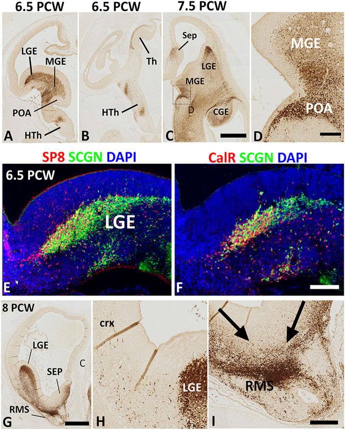
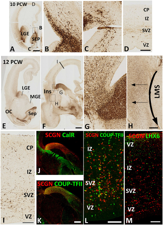
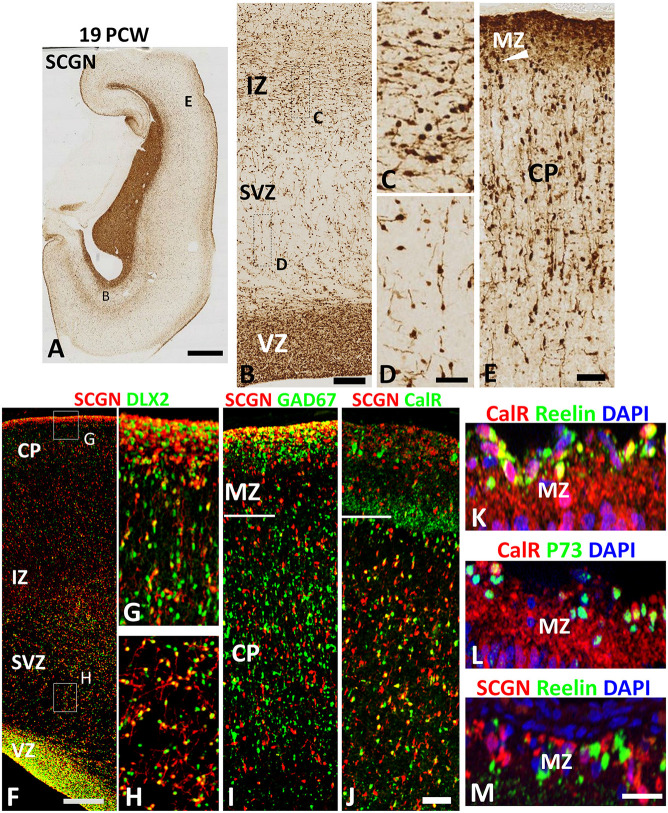
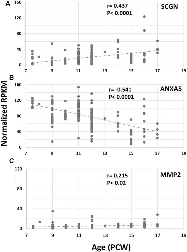
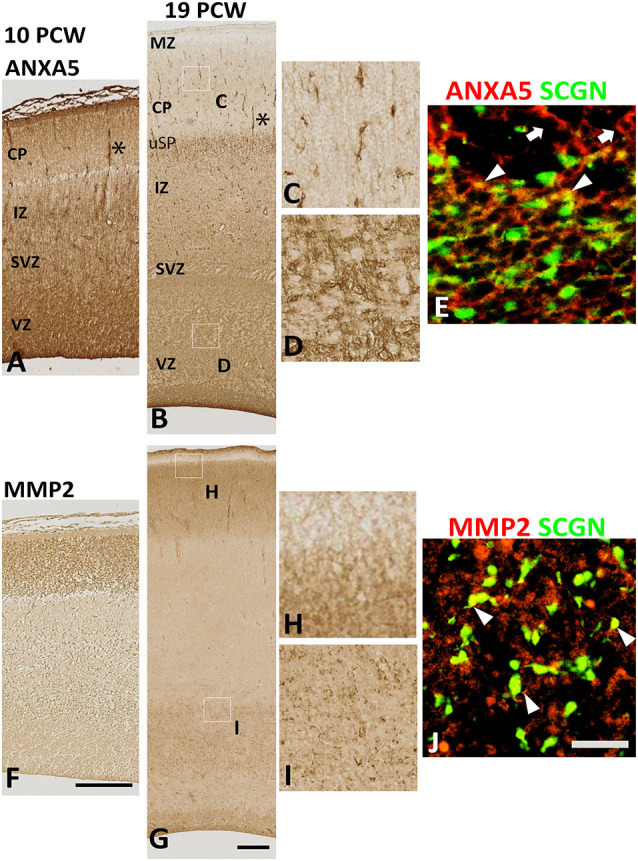
Secretagogin (SCGN) which acts as a calcium signaling sensor, has previously been shown to be expressed by a substantial population of cortical GABAergic neurons at mid-gestation in humans but not in mice. The present study traced SCGN expression in cortical GABAergic neurons in human fetal forebrain from earlier stages than previously studied. Multiple potential origins of SCGN-expressing neurons were identified in the caudal ganglionic eminence (CGE) lateral ganglionic eminence (LGE) septum and preoptic area; these cells largely co-expressed SP8 but not the medial ganglionic eminence marker LHX6. They followed various migration routes to reach their target regions in the neocortex, insular and olfactory cortex (OC) and olfactory bulbs. A robust increase in the number of SCGN-expressing GABAergic cortical neurons was observed in the midgestational period; 58% of DLX2+ neurons expressed SCGN in the cortical wall at 19 post-conceptional weeks (PCW), a higher proportion than expressed calretinin, a marker for GABAergic neurons of LGE/CGE origin. Furthermore, although most SCGN+ neurons co-expressed calretinin in the cortical plate (CP) and deeper layers, in the marginal zone (MZ) SCGN+ and calretinin+ cells formed separate populations. In the adult mouse, it has previously been shown that in the rostral migratory stream (RMS), SCGN, annexin V (ANXA5), and matrix metalloprotease 2 (MMP2) are co-expressed forming a functioning complex that exocytoses MMP2 in response to calcium. In the present study, ANXA5 showed widespread expression throughout the cortical wall, although MMP2 expression was very largely limited to the CP. We found co-expression of these proteins in some SCGN+ neurons in the subventricular zones (SVZ) suggesting a limited role for these cells in remodeling the extracellular matrix, perhaps during cell migration.

Keywords: annexin V; cerebral cortex; ganglionic eminences; inhibitory interneurons; matrix metalloprotease 2; preoptic area; secretagogin; septum.


Figures

Figure 1
Figure 1
Secretagogin (SCGN) expression 6.5–8 post-conceptional weeks (PCW). (A) Rostral coronal section of 6.5 PCW brain; intense SCGN expression in ganglionic eminences (GE) preoptic area (POA) basal telencephalon and hypothalamus (HTh) and (B) Caudal thalamus (Th) and several locations in the hypothalamus. (C) Horizontal section of 7.5 PCW brain; SCGN immunoreactivity in the medial ganglionic eminence (MGE), lateral ganglionic eminence (LGE), caudal ganglionic eminence (CGE), septum (SEP), and POA. (D) A high magnification view of the boxed area in (C) shows many SCGN+ cells that appeared to enter MGE from POA. (E) Double labeling for SCGN and specificity protein 8 (SP8) in LGE showing most SCGN+ cells were double-labeled with SP8, although there is a population of SP8+ only cells in the dorsal most LGE. (F) Double labeling for SCGN and calretinin (CalR) in LGE showing only a proportion of SCGN+ cells co-expressed CalR. (G–I) The rostral coronal section at 8 PCW showing intense SCGN expression in LGE, septum, and rostral migratory stream (RMS, G). Few SCGN+ cells enter cortex from LGE (H). Cells from LGE and septum appeared to be mainly migrating into RMS at this stage (I). Scale bars: 1 mm in (C) (and for A,B) and (G), 50 μm in (D,F) (and for E), and (I) (and for H).
Figure 2
Figure 2
SCGN expression 10–12 PCW. At 10 PCW (A) high expression observed in LGE and SEP. Higher magnification insets show the migration of SCGN+ cells from septum into dorsomedial (B) and ventromedial (C) cortex. In dorsal cortex (D) a small number of SCGN+ cells seen predominantly migrating through subventricular zone (SVZ). By 12 PCW (E,F), SCGN+ expression concentrated in dorsal LGE and low in MGE with higher magnification inset showing SCGN+ cells crossing the pallial/subpallial border to invade lateral cortex (G) and also migrating ventrolaterally in the lateral migratory stream (LMS) towards olfactory cortex (OC) and amygdala (E,F). Some SCGN+ exit LMS to populate insula (Ins, F,H). In the dorsal cortex (I) there are increased numbers of SCGN+ cells compared to 10 PCW and throughout the cortical wall while still favoring the SVZ. A proportion of SCGN+ cells in the LGE co-express CalR (J) but a higher number still co-express COUP-TFII both in the LGE (K) and the cortical wall (L). However, no co-expression with LHX6 observed (M). Scale bars: 1 mm in (A) (and for E); 50 μm in (D) (and for B,C); in (H,I,L,M), and in (K) (and for J).
Figure 3
Figure 3
SCGN expression at 19 PCW. (A) The coronal section at 19 PCW with intense SCGN expression in CGE but also in ventricular zone (VZ) of the cortical wall (B) and in the marginal zone (MZ, E) otherwise moderate expression throughout the cortical wall. Panels (B–D) show that SCGN+ cells exhibit morphology of predominantly tangentially migrating cells in intermediate zone (IZ) but are sparser in SVZ, unlike 10–12 PCW, and show predominantly radial migratory morphology. In the cortical plate (CP) SCGN+ cells also have predominantly radial migratory morphology (E). (F) A high proportion of SCGN+ neurons co-expressed DLX2 in both CP/MZ (G) and SVZ (H). SCGN+ neurons in MZ, in particular, strongly co-expressed GAD67 (I). Co-expression with CalR was abundant in the CP but in MZ, CalR and SCGN showed largely separate patterns of expression (J). In the outer, subgranular layer of the MZ, CalR primarily co-localized with markers of Cajal Retzius cells, Reelin and P73 (K,L) whereas SCGN did not (M). Scale bars: 2 mm in (A); 100 μm in (B); 50 μm in (D) (and for C) (F,J) (and for G–I) and in (M) (and for L,K).
Figure 4
Figure 4
RNAseq for cortical expression of SCGN, ANXA5 and MMP2, 7.5–17 PCW. Changes of expression (normalized RPKM) with age. SCGN expression showed steady and statistically significant increases with age (A) whereas ANXA5 showed a significant decrease in expression (B). MMP2 was modestly expressed at all ages studied but expression increased to a small but significant degree with age (C). Correlation coefficients (r) and p-values for r (p) are displayed on the charts.
Figure 5
Figure 5
Expression of ANXA5 and MMP2 in the cortical wall. (A) At 10 PCW there was strong immunoreactivity for ANXA5 throughout cortical wall especially in MZ, VZ, and blood vessels (*). (B) MMP2 expression was entirely confined to CP. (C) By 19 PCW, ANXA5 expression was still strong in VZ, SVZ (E), and blood vessels (*) but much reduced in MZ. However, many positive cells present in the upper subplate (uSP). Occasional cells with neuronal morphology in CP were also strongly immunostained for ANXA5 (D). In SVZ, both SCGN+ (arrowheads) and SCGN− (arrows) cells expressed ANXA5 (F). MMP2 expression was largely confined to CP (but not MZ, see G,H) but there was also expressed in the VZ and SVZ (G,I) where MMP2 immunoreactivity was found in close apposition to SCGN+ cells (arrowhead, J). MZ, marginal zone; CP, cortical plate; pSP, presubplate; uSP, upper subplate; lSP, lower subplate; IZ, intermediate zone; SVZ, subventricular zone; VZ, ventricular zone. Scale bars: 100 μm in (F) (and for A) in (G) (and for B); 50 μm in (J) (and for C–E, H and I).

References

    1. Alzu’bi A., Clowry G. J. (2019). Expression of ventral telencephalon transcription factors ASCL1 and DLX2 in the early fetal human cerebral cortex. J. Anat. 235, 555–568. 10.1111/joa.12971 - DOI - PMC - PubMed
    1. Alzu’bi A., Homman-Ludiye J., Bourne J. A., Clowry G. J. (2019). Thalamocortical afferents innervate the cortical subplate much earlier in development in primate than in rodent. Cereb. Cortex 29, 1706–1718. 10.1093/cercor/bhy327 - DOI - PMC - PubMed
    1. Alzu’bi A., Lindsay S., Kerwin J., Looi S. J., Khalil F., Clowry G. J. (2017). Distinct cortical and sub-cortical neurogenic domains for GABAergic interneuron precursor transcription factors NKX2. 1, OLIG2 and COUP-TFII in early fetal human telencephalon. Brain Struct. Funct. 222, 2309–2328. 10.1007/s00429-016-1343-5 - DOI - PMC - PubMed
    1. Anders S., Pyl T. P., Huber W. (2014). HTSeq—a Python framework to work with high-throughput sequencing data. Bioinformatics 31, 166–169. 10.1093/bioinformatics/btu638 - DOI - PMC - PubMed
    1. Asgarian Z., Magno L., Ktena N., Harris K. D., Kessaris N. (2019). Hippocampal CA1 somatostatin interneurons originate in the embryonic MGE/POA. Stem Cell Reports 13, 793–802. 10.1016/j.stemcr.2019.09.008 - DOI - PMC - PubMed