

The Role of Oxytocin in Cardiovascular Protection
- PMID: 32982875
- PMCID: PMC7477297
- DOI: 10.3389/fpsyg.2020.02139
The Role of Oxytocin in Cardiovascular Protection
Abstract
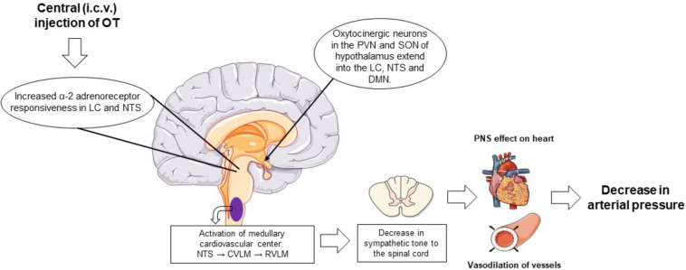
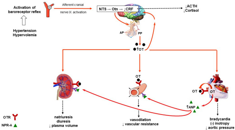
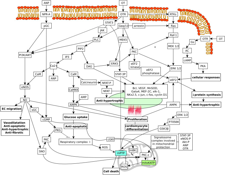
The beneficial effects of oxytocin on infarct size and functional recovery of the ischemic reperfused heart are well documented. The mechanisms for this cardioprotection are not well defined. Evidence indicates that oxytocin treatment improves cardiac work, reduces apoptosis and inflammation, and increases scar vascularization. Oxytocin-mediated cytoprotection involves the production of cGMP stimulated by local release of atrial natriuretic peptide and synthesis of nitric oxide. Treatment with oxytocin reduces the expression of proinflammatory cytokines and reduces immune cell infiltration. Oxytocin also stimulates differentiation stem cells to cardiomyocyte lineages as well as generation of endothelial and smooth muscle cells, promoting angiogenesis. The beneficial actions of oxytocin may include the increase in glucose uptake by cardiomyocytes, reduction in cardiomyocyte hypertrophy, decrease in oxidative stress, and mitochondrial protection of several cell types. In cardiac and cellular models of ischemia and reperfusion, acute administration of oxytocin at the onset of reperfusion enhances cardiomyocyte viability and function by activating Pi3K and Akt phosphorylation and downstream cellular signaling. Reperfusion injury salvage kinase and signal transducer and activator of transcription proteins cardioprotective pathways are involved. Oxytocin is cardioprotective by reducing the inflammatory response and improving cardiovascular and metabolic function. Because of its pleiotropic nature, this peptide demonstrates a clear potential for the treatment of cardiovascular pathologies. In this review, we discuss the possible cellular mechanisms of action of oxytocin involved in cardioprotection.
Keywords: atrial natriuretic peptide; cardiomyocyte; heart; oxytocin; oxytocin—therapeutic use.
Copyright © 2020 Jankowski, Broderick and Gutkowska.
Figures
Similar articles
-
The role of oxytocin in cardiovascular regulation.Braz J Med Biol Res. 2014 Feb;47(3):206-14. doi: 10.1590/1414-431X20133309. Epub 2014 Mar 18. Braz J Med Biol Res. 2014. PMID: 24676493 Free PMC article. Review.
-
Molecular mechanisms underlying oxytocin-induced cardiomyocyte protection from simulated ischemia-reperfusion.Mol Cell Endocrinol. 2015 Sep 5;412:170-81. doi: 10.1016/j.mce.2015.04.028. Epub 2015 May 8. Mol Cell Endocrinol. 2015. PMID: 25963797
-
cGMP-Elevating Compounds and Ischemic Conditioning Provide Cardioprotection Against Ischemia and Reperfusion Injury via Cardiomyocyte-Specific BK Channels.Circulation. 2017 Dec 12;136(24):2337-2355. doi: 10.1161/CIRCULATIONAHA.117.028723. Epub 2017 Oct 19. Circulation. 2017. PMID: 29051185
-
Oxytocin revisited: its role in cardiovascular regulation.J Neuroendocrinol. 2012 Apr;24(4):599-608. doi: 10.1111/j.1365-2826.2011.02235.x. J Neuroendocrinol. 2012. PMID: 21981277 Review.
-
The SAFE pathway is involved in the postconditioning mechanism of oxytocin in isolated rat heart.Peptides. 2019 Jan;111:142-151. doi: 10.1016/j.peptides.2018.04.002. Epub 2018 Apr 7. Peptides. 2019. PMID: 29635063
Cited by
-
Oxytocin Downregulates the CaV1.2 L-Type Ca2+ Channel via Gi/cAMP/PKA/CREB Signaling Pathway in Cardiomyocytes.Membranes (Basel). 2021 Mar 25;11(4):234. doi: 10.3390/membranes11040234. Membranes (Basel). 2021. PMID: 33806201 Free PMC article.
-
Oxytocin Receptor Expression and Activation in Parasympathetic Brainstem Cardiac Vagal Neurons.eNeuro. 2025 Aug 22;12(8):ENEURO.0204-25.2025. doi: 10.1523/ENEURO.0204-25.2025. Print 2025 Aug. eNeuro. 2025. PMID: 40769582 Free PMC article.
-
The Long Way of Oxytocin from the Uterus to the Heart in 70 Years from Its Discovery.Int J Mol Sci. 2023 Jan 29;24(3):2556. doi: 10.3390/ijms24032556. Int J Mol Sci. 2023. PMID: 36768879 Free PMC article. Review.
-
Love and longevity: A Social Dependency Hypothesis.Compr Psychoneuroendocrinol. 2021 Sep 30;8:100088. doi: 10.1016/j.cpnec.2021.100088. eCollection 2021 Nov. Compr Psychoneuroendocrinol. 2021. PMID: 35757670 Free PMC article. Review.
-
Fantastic voyage: Chasing oxytocin from the bedside to the bench and back again.Compr Psychoneuroendocrinol. 2023 Nov 4;17:100213. doi: 10.1016/j.cpnec.2023.100213. eCollection 2024 Feb. Compr Psychoneuroendocrinol. 2023. PMID: 38482487 Free PMC article. Review.
References
-
- Alizadeh A. M., Faghihi M., Khori V., Sohanaki H., Pourkhalili K., Mohammadghasemi F., et al. (2012). Oxytocin protects cardiomyocytes from apoptosis induced by ischemia-reperfusion in rat heart: role of mitochondrial ATP-dependent potassium channel and permeability transition pore. Peptides 36 71–77. 10.1016/j.peptides.2012.03.023 - DOI - PubMed
-
- Alizadeh A. M., Faghihi M., Sadeghipour H. R., Mohammadghasemi F., Imani A., Houshmand F., et al. (2010). Oxytocin protects rat heart against ischemia-reperfusion injury via pathway involving mitochondrial ATP-dependent potassium channel. Peptides 31 1341–1345. 10.1016/j.peptides.2010.04.012 - DOI - PubMed
Publication types
LinkOut - more resources
Full Text Sources