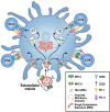
Infectious Tolerance as Seen With 2020 Vision: The Role of IL-35 and Extracellular Vesicles
- PMID: 32983104
- PMCID: PMC7480133
- DOI: 10.3389/fimmu.2020.01867
Infectious Tolerance as Seen With 2020 Vision: The Role of IL-35 and Extracellular Vesicles
Abstract
Originally identified as lymphocyte regulation of fellow lymphocytes, our understanding of infectious tolerance has undergone significant evolutions in understanding since being proposed in the early 1970s by Gershon and Kondo and expanded upon by Herman Waldman two decades later. The evolution of our understanding of infectious tolerance has coincided with significant cellular and humoral discoveries. The early studies leading to the isolation and identification of Regulatory T cells (Tregs) and cytokines including TGFβ and IL-10 in the control of peripheral tolerance was a paradigm shift in our understanding of infectious tolerance. More recently, another potential, paradigm shift in our understanding of the "infectious" aspect of infectious tolerance was proposed, identifying extracellular vesicles (EVs) as a mechanism for propagating infectious tolerance. In this review, we will outline the history of infectious tolerance, focusing on a potential EV mechanism for infectious tolerance and a novel, EV-associated form for the cytokine IL-35, ideally suited to the task of propagating tolerance by "infecting" other lymphocytes.
Keywords: IL-35; exosomes; extracellular vesicle; immunotherapy; infectious tolerance.
Copyright © 2020 Sullivan, AlAdra, Olson, McNeel and Burlingham.
Figures


Similar articles
-
Treg-Cell-Derived IL-35-Coated Extracellular Vesicles Promote Infectious Tolerance.Cell Rep. 2020 Jan 28;30(4):1039-1051.e5. doi: 10.1016/j.celrep.2019.12.081. Cell Rep. 2020. PMID: 31995748 Free PMC article.
-
T regulatory cells-derived extracellular vesicles and their contribution to the generation of immune tolerance.J Leukoc Biol. 2020 Sep;108(3):813-824. doi: 10.1002/JLB.3MR0420-533RR. Epub 2020 Jun 12. J Leukoc Biol. 2020. PMID: 32531824 Review.
-
Glioma-derived extracellular vesicles selectively suppress immune responses.Neuro Oncol. 2016 Apr;18(4):497-506. doi: 10.1093/neuonc/nov170. Epub 2015 Sep 18. Neuro Oncol. 2016. PMID: 26385614 Free PMC article.
-
Self-tolerance revisited.Stud Hist Philos Biol Biomed Sci. 2016 Feb;55:128-32. doi: 10.1016/j.shpsc.2015.11.006. Stud Hist Philos Biol Biomed Sci. 2016. PMID: 27200443 No abstract available.
-
Special regulatory T-cell review: A rose by any other name: from suppressor T cells to Tregs, approbation to unbridled enthusiasm.Immunology. 2008 Jan;123(1):20-7. doi: 10.1111/j.1365-2567.2007.02779.x. Immunology. 2008. PMID: 18154615 Free PMC article. Review.
Cited by
-
Regulatory T Cell-Enhancing Therapies to Treat Atherosclerosis.Cells. 2021 Mar 24;10(4):723. doi: 10.3390/cells10040723. Cells. 2021. PMID: 33805071 Free PMC article. Review.
-
IL-27-containing exosomes secreted by innate B-1a cells suppress and ameliorate uveitis.Front Immunol. 2023 Jun 2;14:1071162. doi: 10.3389/fimmu.2023.1071162. eCollection 2023. Front Immunol. 2023. PMID: 37334383 Free PMC article.
-
Interleukin 35-producing B cells prolong the survival of GVHD mice by secreting exosomes with membrane-bound IL-35 and upregulating PD-1/LAG-3 checkpoint proteins.Theranostics. 2025 Feb 25;15(8):3610-3626. doi: 10.7150/thno.105069. eCollection 2025. Theranostics. 2025. PMID: 40093899 Free PMC article.
-
Immunosuppressive Mechanisms of Regulatory B Cells.Front Immunol. 2021 Apr 29;12:611795. doi: 10.3389/fimmu.2021.611795. eCollection 2021. Front Immunol. 2021. PMID: 33995344 Free PMC article. Review.
-
Interleukin-35 pathobiology in periodontal disease: a systematic scoping review.BMC Oral Health. 2021 Mar 20;21(1):139. doi: 10.1186/s12903-021-01515-1. BMC Oral Health. 2021. PMID: 33743678 Free PMC article.
References
-
- Sakaguchi S, Sakaguchi N, Asano M, Itoh M, Toda M. Immunologic self-tolerance maintained by activated T cells expressing IL-2 receptor alpha-chains (CD25). Breakdown of a single mechanism of self-tolerance causes various autoimmune diseases. J Immunol. (1995) 155:1151–64. - PubMed
Publication types
MeSH terms
Substances
Grants and funding
LinkOut - more resources
Full Text Sources

