Case Reports
. 2020 Sep 1:11:2146.
doi: 10.3389/fimmu.2020.02146. eCollection 2020.

Zika Induces Human Placental Damage and Inflammation

Affiliations
Case Reports

Zika Induces Human Placental Damage and Inflammation

Kíssila Rabelo et al. Front Immunol. .

Abstract

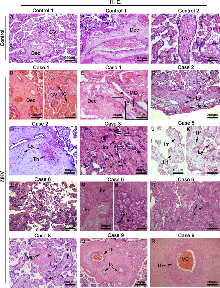
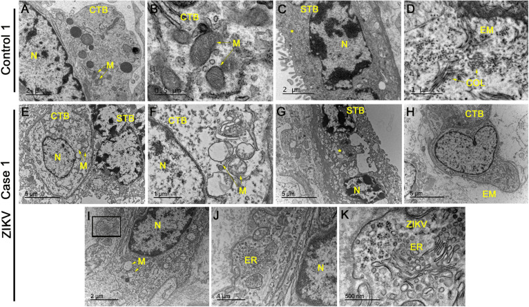
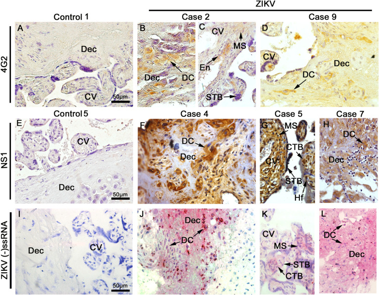
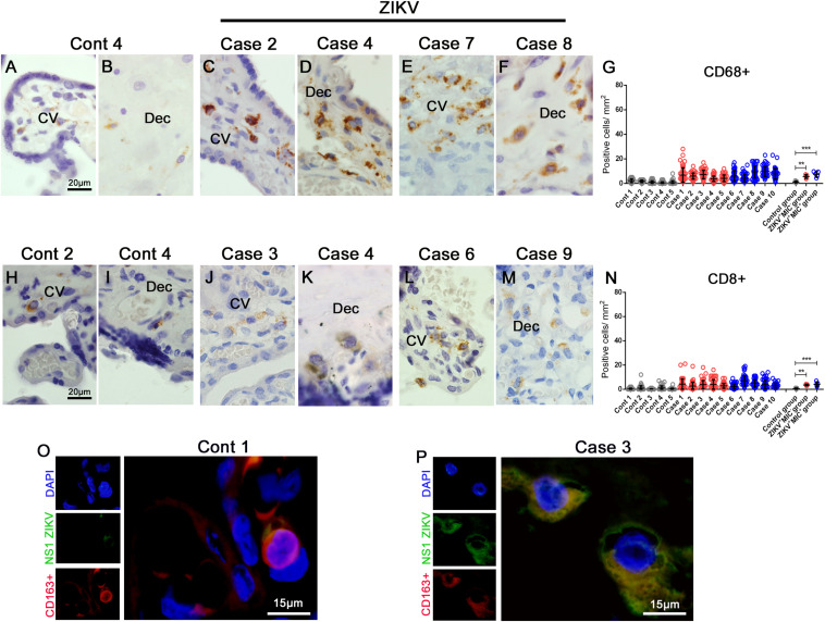
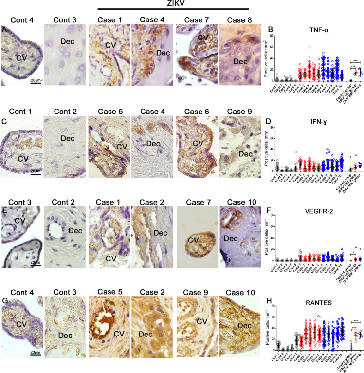
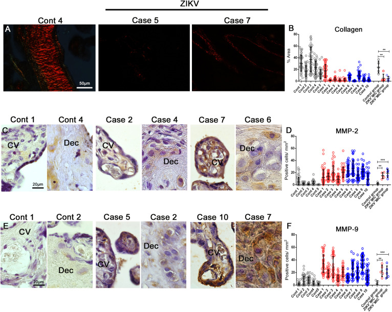
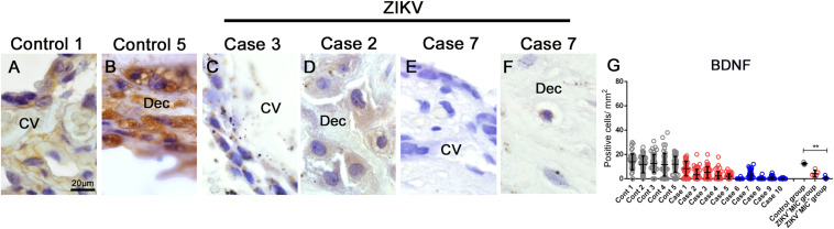
In Brazil, an epidemic of Zika virus (ZIKV) infections was declared in 2015 that coincided with alarming reports of microcephaly in newborns associated with mother infection. Although the virus has placental tropism, changes in the tissue morphology and immunity of infected patients have not yet been elucidated. Here, we investigated the histopathological and ultrastructural changes along with the immunological profile and the BDNF expression in rare placental material. Tissues were obtained in the 2015-2016 Brazilian epidemic, of ten ZIKV-infected patients during pregnancy, five resulting in cases of fetal microcephaly and five non-microcephaly, compared to five non-infected control placentae. Viral antigens were only detected in samples from the ZIKV infected patients. Infected placentae presented histopathological severe damage, while the ultrastructural evaluation showed abnormal organelles, such as clusters of virus-like particles consistent with the ZIKV dimensions. Increased infiltration of CD68+ and TCD8+ cells, expression of MMPs, cytokines (IFN-γ and TNF-α) and other immunological mediators (RANTES/CCL5 and VEGFR-2) confirmed excessive inflammation and vascular permeability dysfunction. An evaluation of BDNF showed a decrease that could modulate neuronal damage in the developing fetus. The placental changes caused by ZIKV are not pathognomonic, however, the data provide evidence that this infection leads to severe placental injury.

Keywords: cytokines; flavivirus; histopathology; immune response; ultrastructure.


Figures

FIGURE 1
FIGURE 1
Placentae histopathology. (A–C) Placentae from non-ZIKV patients stained with H&E and presenting normal features: maternal decidua (Dec) and chorionic villi (CV). (D–R) Placentae from ZIKV infected patients that presented a range of different alterations such as VC, vascular congestion; IC, intervillous congestion; MØ, macrophage infiltrate; Ly, lymphocytic infiltrate villous; He, hemorrhage; Th, endothelial thickening; Im, immature chorionic villi; Hf, Hofbauer cells; Ca, calcification; Fi, fibrin areas; IFT, infarct, and Is, ischemia.
FIGURE 2
FIGURE 2
Electron microscopy analysis of placental sections showed alterations and virus-like particles in ZIKV infected samples. (A–D) Electron microscopy images of ultrathin sections of placental tissue from a single, non-ZIKV infected mother that exhibited regular cytotrophoblasts (CTB), syncytiotrophoblasts (STB), nucleus (N), mitochondria (M), and collagen filament structures (COL). (E–H) Electron micrographs of ultrathin sections of placental tissue from different ZIKV-infected mothers showing CTB with alterations in the cytoplasm, nuclear variation (N) and swollen mitochondria with a loss of cristae and membrane rupture. The identified STB presented an enlargement of vesicles and apoptotic bodies (asterisks) along with an absence of normal membrane extensions and evidence of microvesicle secretion. The extracellular matrix (EM) did not present collagen filaments. (I–K) Identification of clusters of virus-like particles that were positioned near the endoplasmic reticulum (ER) of CTB and ∼25 nm in diameter, which is consistent with the dimensions of ZIKV.
FIGURE 3
FIGURE 3
Detection of viral antigens. Tissue sections were probed by immunohistochemistry for the E (A–D) and NS1 (E–H) antigens of ZIKV as well as by in situ hybridization for the genome of ZIKV (I–L). Representative images of control placentae (A,E,I) showing the absence of reactivity for the antigens and genome of ZIKV. Representative images of placenta from ZIKV+ placenta, independent of the cephalic circumference of the infant, show: (B–D) the presence of E protein in decidual cells (DC) of decidua (Dec), mesenchymal cells (MS), endothelial cells (En), and syncytiotrophoblast (STB) of chorionic villi (CV) that was independent of the cephalic circumference of the infant; (F–H) NS1 protein detected in decidual cells (DC) of decidua (Dec), mesenchymal cells (MS), Hofbauer (Hf), syncytiotrophoblast (STB) and cytotrophoblast (CTB) of chorionic villi (CV); and (J–L) Detection of ZIKV RNA negative strand by in situ hybridization in decidual cells (DC) of decidua (Dec), mesenchymal cells (MS), syncytiotrophoblast (STB) and cytotrophoblast (CTB) of chorionic villi (CV).
FIGURE 4
FIGURE 4
Increased cellularity of mononuclear cell subpopulations in ZIKV-infected placental tissues. CD68 and CD8 were detected in placenta samples by immunohistochemistry. (A–B) CD68+ cells in decidua and chorionic villi of control placenta. (C–F) CD68+ cells in decidua and chorionic villi of ZIKV infected placentae. (G) Quantification of CD68+ cells showing a 5 or 6-fold increase in placentae of ZIKV+MIC and ZIKV+MIC+ groups, respectively. (H,I) CD8+ cells in decidua and chorionic villi of control placentae. (J–M) CD8+ cells in decidua and chorionic villi of ZIKV infected placentae. (N) Quantification of CD8+ cells showing a 7 or 8-fold increase in the tissues of ZIKV+MIC and ZIKV+MIC+ groups, respectively. (O,P) Immunofluorescence for the presence of ZIKV NS1 protein (green) and CD163 (red; biomarker for macrophages) showing colocalization. Nuclei were stained using DAPI (blue). (O) ZIKV NS1 antigen was not detected in the control placenta. Data are represented as mean ± SDM. Asterisks indicate differences that are statistically significant between groups (***p < 0.001).
FIGURE 5
FIGURE 5
Cytokine-producing cell profile. Detection of TNF-α, IFN-γ, VEGFR-2 and RANTES/CCL5 by immunohistochemistry show (A) TNF-α in cells of chorionic villi in control placentae (Left panel) and ZIKV infected placentae (Right panel). (C) Production of IFN-γ in macrophages as well as endothelial cells in chorionic villi and decidual cells of the decidua of control placentae (Left panel) and ZIKV infected placentae (Right panel). (E) VEGFR-2 was expressed in endothelial cells of decidua and chorionic villi in control placentae (Left panel) and ZIKV infected placentae (Right panel). (G) RANTES/CCL5 present mainly in the endothelium and Hofbauer cells located within the chorionic villi and decidual cells and syncytiotrophoblasts of the decidua in control placentae (Left panel) and ZIKV infected placentae (Right panel). Quantification of the cells positive for (B,D,F,H) Quantification of the number of cells expressing TNF-α (B), IFN-γ (D), VEGFR2 (F) and RANTES/CCL5 (H) showed an increased expression of local pro-inflammatory cytokines and mediators in ZIKV positive placentae compared to controls. Data are represented as mean ± SDM. Asterisks indicate differences that are statistically significant between groups (**p < 0.01) or (***p < 0.001).
FIGURE 6
FIGURE 6
Detection and quantification of collagen, MMP-2 and MMP-9 collagenases expression. (A) Collagen detection by Picro Sirius Red staining in placental tissues. (B) The percent collagen area was quantified in all cases that showed a decrease in the expression of collagen in infected placentae. (C–E) Detection of MMP-2 and MMP-9 in decidual cells and cells located within the chorionic villi in both control and ZIKV infected placentae. (D–F) Quantification of the number of cells expressing MMP-2 and MMP-9 showed an increased expression in ZIKV infected placental tissues. Data are represented as mean ± SDM. Asterisks indicate differences that are statistically significant between groups (**p < 0.01) or (***p < 0.001).
FIGURE 7
FIGURE 7
BDNF detection and quantification in placental tissues. (A–F) Detection of BDNF in cells of chorionic villi and maternal decidua of controls and ZIKV infected placentae by immunohistochemistry. (G) A quantification of the number of cells expressing BDNF showed a decreased in the expression of this hormone in ZIKV+ MIC+ cases. Data are represented as mean ± SDM. Asterisks indicate differences that are statistically significant between groups (**p < 0.01).

References

    1. Musso D, Ko AI, Baud D. Zika virus infection – after the pandemic. N Engl J Med. (2019) 381:1444–57. 10.1056/NEJMra1808246 - DOI - PubMed
    1. Faria NR, Do Socorro Da Silva Azevedo R, Kraemer MUG, Souza R, Cunha MS, Hill SC, et al. Zika virus in the Americas: early epidemiological and genetic findings. Science. (2016) 352:345–9. 10.1126/science.aaf5036 - DOI - PMC - PubMed
    1. Dyer O. Zika virus spreads across Americas as concerns mount over birth defects. BMJ. (2015) 351:h6983. 10.1136/bmj.h6983 - DOI - PubMed
    1. Rabelo K, de Souza Campos Fernandes RC, de Souza LJ, de Souza TL, dos Santos FB, Nunes PCG, et al. Placental histopathology and clinical presentation of severe congenital Zika syndrome in a human immunodeficiency virus-exposed uninfected infant. Front Immunol. (2017) 8:1704. 10.3389/fimmu.2017.01704 - DOI - PMC - PubMed
    1. Araujo AQCA, Silva MTT, Araujo APQC. Zika virus-associated neurological disorders: a review. Brain. (2016) 139:2122–30. 10.1093/brain/aww158 - DOI - PubMed

Publication types

MeSH terms