Review
. 2020 Sep 10:16:2260-2271.
doi: 10.3762/bjoc.16.187. eCollection 2020.

Tools for generating and analyzing glycan microarray data

Affiliations
Review

Tools for generating and analyzing glycan microarray data

Akul Y Mehta et al. Beilstein J Org Chem. .

Abstract

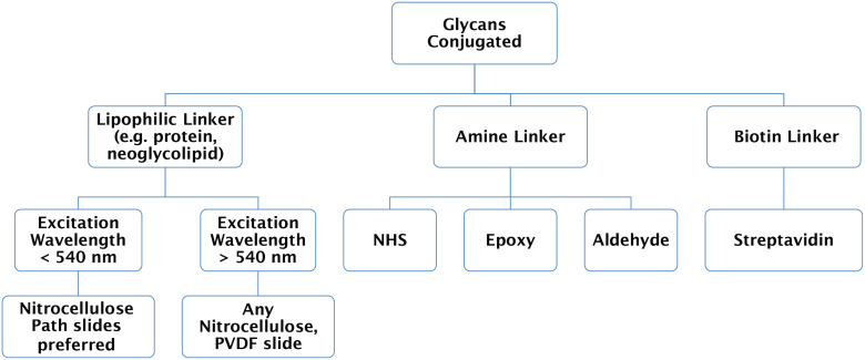
Glycans are one of the major biological polymers found in the mammalian body. They play a vital role in a number of physiologic and pathologic conditions. Glycan microarrays allow a plethora of information to be obtained on protein-glycan binding interactions. In this review, we describe the intricacies of the generation of glycan microarray data and the experimental methods for studying binding. We highlight the importance of this knowledge before moving on to the data analysis. We then highlight a number of tools for the analysis of glycan microarray data such as data repositories, data visualization and manual analysis tools, automated analysis tools and structural informatics tools.

Keywords: data analysis; glycan binding; glycan microarray; glycomics; informatics.


Figures

Figure 1
Figure 1
Overview of a typical glycan microarray workflow, beginning with the obtention of glycans to analysis of binding data. Briefly, glycans are chemically or enzymatically synthesized, or isolated and purified from either source materials, and then conjugated with a linker which is appropriate for the printing surface. The glycoconjugates are then printed upon appropriately functionalized slides, followed by blocking; the printed slides are stored under ideal conditions prior to experiments. Many arrays can be printed on a single slide, termed sub-arrays. The slides can then be used in a glycan microarray experiment where they are incubated with a glycan binding protein (GBP), such as lectin, antibody, or serum, virus, etc., followed by addition of a detection reagent, if the primary analyte was not fluorescently labeled, for example a fluorescent secondary antibody or streptavidin. After washing the slide to remove unbound material, the bound material is then identified and measured by scanning using a fluorescence microarray scanner. The image produced can then be analyzed using automated or manual methods to generate the array results. These results can in turn be stored in a database, or interpreted either manually or by automatic algorithms. The glycan structures in the figure were produced using GlycoGlyph [29].
Figure 2
Figure 2
A decision tree to determine which type of surface to use depending on glycoconjugate and linker type, and scanning requirements. These linker types and surfaces are the most commonly used and there are more specialized surfaces and linker chemistries that are commercially available.
Figure 3
Figure 3
Screenshot of microarray databases: (A) Screenshot of an example of CFG glycan array data; (B) screenshot of an example of Imperial College microarray data online portal.
Figure 4
Figure 4
A demonstration of glycan array data visualization with GLAD. The dataset used is from Byrd-Leotis et al. [16], and is provided as a GLAD session file in the manuscript. The dataset contains data on the CFG microarray for various drift H3N2 strains of influenza (#1–10) in comparison to the distinct New York H3N2 strain (#11) and two H1N1 strains (#12 and #13). Details regarding the strains and the experiment can be found in the original paper. The legend for the colors is provided at the bottom of the figure. (A) The complete dataset can be visualized using the force graph where each strain of virus is represented by the colored circle, while each glycan is represented by a grey circle. The links between the virus to the glycan nodes are determined by the intensity of binding observed on the microarray, i.e., the higher the intensity the shorter is the link length. A short simulation is run to optimize the distances between the nodes so as to accommodate this link length and as a result of this nodes which bind similar glycans cluster together in comparison to those which bind others. Thus, the drift strains (#1–10) cluster together in the red cluster, in comparison to the New York H3N2 strain in green, all of which are separated from the purple cluster of the H1N1 strains. (B) To visualize specific binding to lactosamine (Galb1-4GalNAc) in the absence of sialic acid, the dataset is filtered for lactosamine containing glycan data and data for glycans with Neu5Ac are eliminated from the pool. A Bubble Plot of this filtered data shows clearly that the drift strains (#1–10) bind better to the lactosamine containing glycans without Neu5Ac as compared to the New York H3N2 strain and the two H1N1 strains. This analysis clearly shows the difference of the drift H3N2 strains which have acquired ability to bind lactosamine containing glycans.

References

    1. Stern R, Jedrzejas M J. Chem Rev. 2008;108:5061–5085. doi: 10.1021/cr078240l. - DOI - PubMed
    1. Colley K J, Varki A, Kinoshita T. Cellular Organization of Glycosylation. In: Varki A, Cummings R D, Esko J D, et al., editors. Essentials of Glycobiology. Cold Spring Harbor, NY, U.S.A.: Cold Spring Harbor Laboratory Press; 2015. pp. 41–49.
    1. Milland J, Sandrin M S. Tissue Antigens. 2006;68:459–466. doi: 10.1111/j.1399-0039.2006.00721.x. - DOI - PubMed
    1. Engle D D, Tiriac H, Rivera K D, Pommier A, Whalen S, Oni T E, Alagesan B, Lee E J, Yao M A, Lucito M S, et al. Science. 2019;364:1156–1162. doi: 10.1126/science.aaw3145. - DOI - PMC - PubMed
    1. Leppänen A, White S P, Helin J, McEver R P, Cummings R D. J Biol Chem. 2000;275(50):39569–39578. doi: 10.1074/jbc.m005005200. - DOI - PubMed

LinkOut - more resources