

[Temporal meningocele and anophtalmia: about a case]
- PMID: 32983326
- PMCID: PMC7501746
- DOI: 10.11604/pamj.2020.37.8.24930
[Temporal meningocele and anophtalmia: about a case]
Abstract
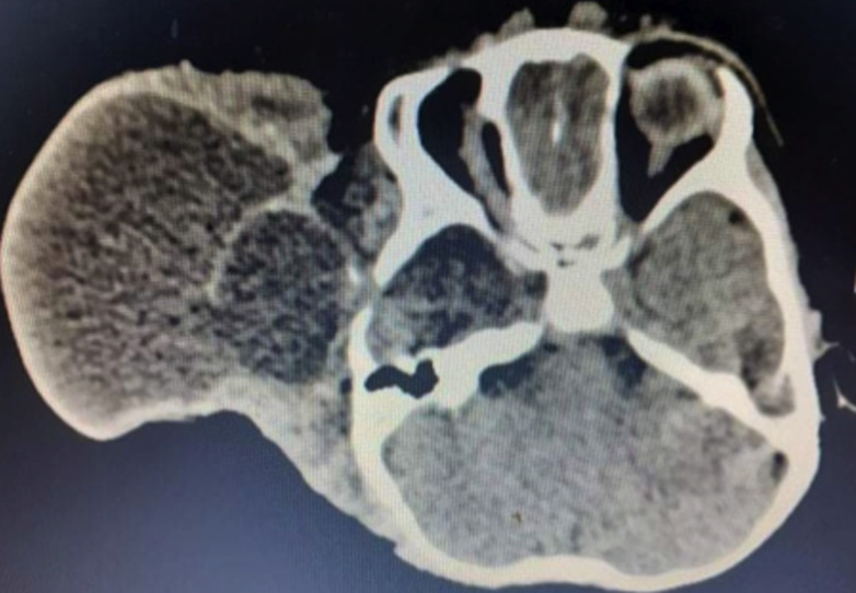
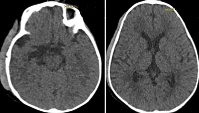
We here report the case of a 12-month old infant with congenital polymalformation including right temporal meningocele and homolateral eyeball aplasia. Brain CT scan confirmed this malformation with bone defect in the right temporal lobe, meningeal hernia containing cerebrospinal fluid and absence of the right eyeball. Surgery was performed to treat meningocele. Patient's outcome was favorable. The purpose of this study was to highlight the rarity of this disease on the basis of a literature review.
Les auteurs rapportent un cas d´un garcon de 12 mois pour une polymalformation congénitale à type de méningocèle temporale droite et une aplasie du globe oculaire homolatérale qui était présent depuis sa naissance. Le scanner cérébral confirmait la malformation avec un défect osseux au niveau temporal droit, une hernie de la méninge contenant du liquide cérébro-spinal et une absence du globe oculaire droit. La chirurgie était pratiquée pour la méningocèle. L´évolution était favorable. Notre objectif est de montrer la rareté de cette affection à la lumière d´une revue de la littérature.
Keywords: Anophthalmia; meningocele; temporal bone.
Copyright: Patrick Rakotozanany et al.
Conflict of interest statement
Les auteurs ne déclarent aucun conflit d´intérêts.
Figures


References
-
- Botto LD, Moore CA, Khoury MJ, Erickson JD. Neural-tube defects. N Engl J Med. 1999 Nov 11;341(20):1509–19. - PubMed
-
- Cataltepe O, Ozcan OE. Bilateral orbital encephaloceles, an unusual cause of exophthalmos. J Clin Neuroophthalmol. 1990 Jun;10(2):131–4. - PubMed
-
- Charoonsmith T. Review of 310 patients with frontoethmoidal mingoencephaloceles. Presented at plastic surgery transactions. VIII Internetional congress of plastic and reconstructive surgery, Montreal, Canada, 198.
-
- David DJ. Cephaloceles: classification, pathology, and management: a review. J Craniofac Surg. 1993 Oct;4(4):192–202. - PubMed
-
- David M, Pourpre H. Neuro-Chirurgie Paris. Flammarion. 1961;973:518–534.
Publication types
MeSH terms
LinkOut - more resources
Full Text Sources
