. 2020 Sep 23:10:113.
doi: 10.1186/s13578-020-00472-6. eCollection 2020.

Vascular NRP2 triggers PNET angiogenesis by activating the SSH1-cofilin axis

Affiliations

Vascular NRP2 triggers PNET angiogenesis by activating the SSH1-cofilin axis

Xi Luo et al. Cell Biosci. .

Abstract

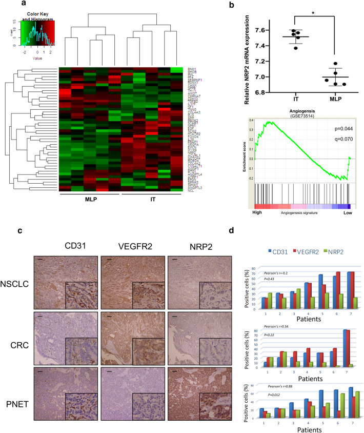
Background: Angiogenesis is a critical step in the growth of pancreatic neuroendocrine tumors (PNETs) and may be a selective target for PNET therapy. However, PNETs are robustly resistant to current anti-angiogenic therapies that primarily target the VEGFR pathway. Thus, the mechanism of PNET angiogenesis urgently needs to be clarified.

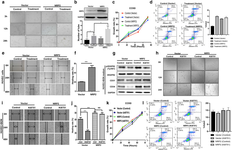
Methods: Dataset analysis was used to identify angiogenesis-related genes in PNETs. Immunohistochemistry was performed to determine the relationship among Neuropilin 2 (NRP2), VEGFR2 and CD31. Cell proliferation, wound-healing and tube formation assays were performed to clarify the function of NRP2 in angiogenesis. The mechanism involved in NRP2-induced angiogenesis was detected by constructing plasmids with mutant variants and performing Western blot, and immunofluorescence assays. A mouse model was used to evaluate the effect of the NRP2 antibody in vivo, and clinical data were collected from patient records to verify the association between NRP2 and patient prognosis.

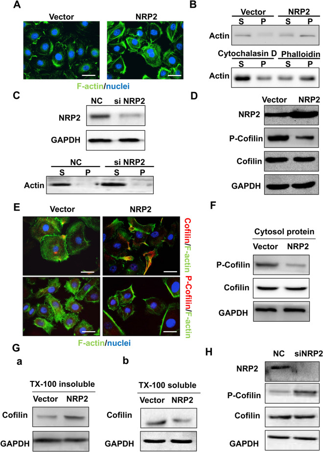
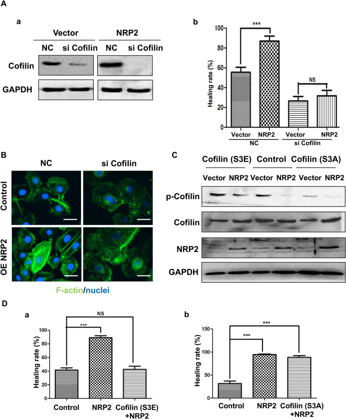
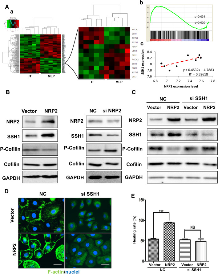
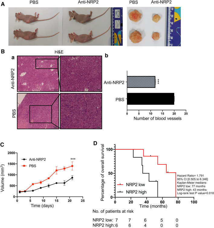
Results: NRP2, a VEGFR2 co-receptor, was positively correlated with vascularity but not with VEGFR2 in PNET tissues. NRP2 promoted the migration of human umbilical vein endothelial cells (HUVECs) cultured in the presence of conditioned medium PNET cells via a VEGF/VEGFR2-independent pathway. Moreover, NRP2 induced F-actin polymerization by activating the actin-binding protein cofilin. Cofilin phosphatase slingshot-1 (SSH1) was highly expressed in NRP2-activating cofilin, and silencing SSH1 ameliorated NRP2-activated HUVEC migration and F-actin polymerization. Furthermore, blocking NRP2 in vivo suppressed PNET angiogenesis and tumor growth. Finally, elevated NRP2 expression was associated with poor prognosis in PNET patients.

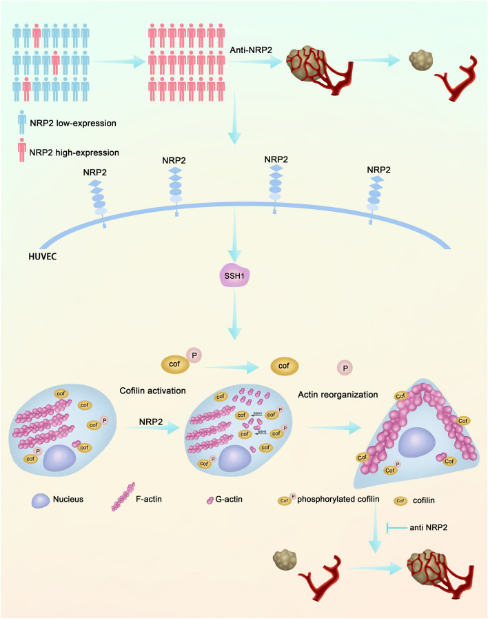
Conclusion: Vascular NRP2 promotes PNET angiogenesis by activating the SSH1/cofilin/actin axis. Our findings demonstrate that NRP2 is an important regulator of angiogenesis and a potential therapeutic target of anti-angiogenesis therapy for PNET.

Keywords: Angiogenesis; Cofilin; Neuropilin 2; Pancreatic neuroendocrine tumor; SSH1.


Conflict of interest statement

Competing interestsThe authors declare that they have no competing interests.

Figures

Fig. 1
Fig. 1
NRP2 expression is positively correlated with PNET vascularity. a Heatmap summarizing the angiogenesis process signature (GSE73514) in metastatic-like primary (MLP) tumor and islet tumor (IT) samples. b Top: Comparison of NRP2 mRNA expression between the MLP and IT groups. Bottom: GSEA mountain plot showing a strong association between the MLP and IT groups. The data are presented as the means ± SD. *P ≤ 0.05 by Student’s t test. C, Representative expression of CD31, VEGFR2 and NRP2 in non-small-cell lung cancer (NSCLC), colorectal cancer (CRC) and pancreatic neuroendocrine tumor (PNET) specimens according to immunohistochemistry assays. d Correlation histograms of CD31, VEGFR2 and NRP2 expression in NSCLC, CRC and PNET specimens according to immunohistochemistry. Each number on the horizontal axis represents one specimen from the patients. The bars in the histograms show the mean percentage of positively staining cells under 5 randomly selected microscopic fields (20x). The Spearman R value and P value in the figures reflect the correlations of NRP2 and CD31 expression
Fig. 2
Fig. 2
NRP2 modulates angiogenesis by promoting HUVEC migration via a VEGF/VEGFR2-independent pathway. a HUVECs were cultured in the presence or absence of conditioned medium from BON cells (treatment and control, respectively) and then transfected with a vector control or NRP2 overexpression plasmid before they were seeded for the capillary tube formation assay. Representative images at 4, 12 and 24 h after plating are shown. b Quantification of the number of complete and broken tubes at 6 h from a representative experiment. Data are shown as the mean ± SD of three independent experiments. *P ≤ 0.05 by Student’s t test. c HUVECs were treated with conditioned medium from BON cells for 24 h and then transfected with a vector or NRP2 overexpression plasmid before they were subjected to a CCK8 assay. d After HUVECs were cultured in the presence or absence of conditioned medium from BON cells (treatment and control, respectively) and transduced with the NRP2-overexpressing plasmid, flow cytometry was performed to assess apoptosis. e Representative images for the wound-healing assay at 0, 24 and 48 h after scratching for the 4 different cell groups (HUVECs with or without NRP2 overexpression cultured in the presence or absence of conditioned medium from BON cells). f Quantification of the healing rate at 48 h after wound-healing assays in HUVECs cultured in the presence or absence of conditioned medium from BON cells followed by transfection with empty vector or NRP2 plasmid. The data are shown as the means ± SD of three independent experiments. ***P ≤ 0.001 by Student’s t test. g HUVECs were transfected with empty vector or an NRP2 overexpression plasmid and then treated with the VEGFR2-specific inhibitor KI8751. Western blotting assays were performed to determine the levels of VEGFR2 phosphorylation at Tyr951 as well as the total protein levels of VEGFR2, CD31, CD34 and GAPDH. h Control and NRP2-overexpressing HUVECs were treated with KI8751 and PBS and evaluated for tube formation. i After HUVECs were cultured in the presence or absence of conditioned medium from BON cells for 24 h, they were transfected with a vector or NRP2 overexpression plasmid. These cells were subsequently treated with PBS (control) or KI8751, and a wound-healing assay was performed. Representative image of three independent experiments is shown. j Qualification of the wound-healing rate at 48 h in HUVEC-vector or HUVEC-NRP2 cells treated with PBS or KI8751. The data are shown as the mean ± SD of three independent experiments. **P ≤ 0.01 by Student’s t test. k After HUVECs were transfected with empty vector or an NRP2 overexpression plasmid, they were treated with PBS or KI8751 and subjected to the CCK8 assay at 24, 48 and 72 h after treatment. l Flow cytometry assay was performed in the 4 cell groups described in Fig. 2k
Fig. 3
Fig. 3
NRP2 induced F-actin polymerization via the active actin-binding protein cofilin. a Immunofluorescence analysis was performed using FITC-labelled phalloidin (F-actin; green), and the nuclei were stained with DAPI (blue). An overlay of the two fluorescent signals is shown (× 1000). b F-actin and G-actin fractions were prepared from HUVECs transfected with empty vector or NRP2 overexpression plasmid (Top). HUVECs were treated with either F-actin depolymerization factor (cytochalasin D) as a positive control or F-actin enhancing factor (phalloidin) as a negative control. The F-actin and G-actin fractions were prepared and subjected to Western blot analysis [S, supernatant fraction (G-actin); P, pellet fraction (F-actin)] (bottom). c HUVECs were transfected with si-control or si-NRP2 and subjected to Western blot analysis using the indicated antibodies. d After HUVECs were transfected with empty vector or an NRP2 overexpression plasmid, they were lysed and subjected to Western blot analysis with the indicated antibodies. e HUVEC-vector and HUVEC-NRP2 cells were coimmunostained with FITC-labelled F-actin and antibodies targeting total and phosphorylated cofilin. The fluorescent signals of cofilin or p-cofilin (red) along with F-actin (blue) are shown (× 1000). f HUVEC-vector and HUVEC-NRP2 cells were lysed using cytosol buffer and subjected to Western blotting with the indicated antibodies. g After HUVECs transfected with empty vector or an NRP2 overexpression plasmid were lysed in Triton X-100 buffer, the insoluble and soluble fractions were subjected to Western blotting. h After NRP2 was knocked down, HUVEC lysates were subjected to Western blot analysis with the indicated antibodies
Fig. 4
Fig. 4
Cofilin activity mediates NRP2-driven HUVEC migration and actin organization. A HUVEC-vector and HUVEC-NRP2 cells were transfected with scramble or cofilin siRNA. a Cells were lysed and subjected to Western blot analysis with cofilin antibodies. b The migratory properties of the cells were analysed by the wound-healing assay (***P ≤ 0.001 by Student’s t test). The data are presented as averages from three independent experiments. B The fluorescent signals of F-actin (green) and nuclei (blue) are shown (× 1000). C HUVEC-vector and HUVEC-NRP2 cells were transfected with cofilin S3A or S3E before they were lysed and subjected to Western blot analysis with the indicated antibodies. D HUVEC-NRP2 cells were transfected with cofilin S3A (a) or cofilin S3E (b). The migratory properties of the cells were analysed by the wound-healing assay. The data are summarized from three independent experiments
Fig. 5
Fig. 5
NRP2 upregulates cofilin activity by increasing SSH1 expression. A Heatmap of an NCBI dataset (GSE73514) of MLP tumors and ITs. Cluster analysis was performed according to an NRP2 transcription level difference of more than fivefold in 2 groups (red indicates high transcription levels, and blue indicates low transcription levels). a Cluster analysis results. b GSEA mountain plot showing a strong association between the MLP and IT groups. c, Correlation analysis of NRP2 and SSH1 transcription levels. B SSH1 expression in HUVEC-vector and HUVEC-NRP2 cells was detected by Western blot analysis (left panels). SSH1 expression in scramble siRNA- or NRP2 siRNA-treated cells was detected by Western blot analysis (right panels). C HUVEC-vector and HUVEC-NRP2 cells were treated with scramble or SSH1 siRNA. Cells were then lysed and subjected to Western blot analysis with the indicated antibodies. D The fluorescent signals of F-actin (green) and nuclei (blue) are shown (× 1000). E The migratory properties of the cells were analysed by the wound-healing assay. The data are presented as averages from 3 independent experiments (***P ≤ 0.001 by Student’s t test)
Fig. 6
Fig. 6
Downregulation of NRP2 suppresses PNET angiogenesis and tumor growth in vivo and correlates with increased survival in patients. a The xenograft experiments in vivo with the mouse model with BON cells. After mice were injected with anti-NRP2 antibody or PBS intraperitoneally, the xenografts were dissected. b H&E staining was performed to determine the number of vessels in the xenograft tumors. The vascular number was calculated as the mean counts of vessels in 5 fields under 20 × magnification. ***P ≤ 0.001 by Student’s t test. c Tumor sizes were measured every other day after injection with PBS or NRP2 antibody. d Survival curve of PNET patients in the high and low NRP2 expression groups
Fig. 7
Fig. 7
Proposed model of vascular NRP2 triggers PNET angiogenesis via activating SSH1-cofilin pathway

References

    1. Singh S, Asa SL, Dey C, Kennecke H, Laidley D, Law C, et al. Diagnosis and management of gastrointestinal neuroendocrine tumors: an evidence-based Canadian consensus. Cancer Treat Rev. 2016;47:32–45. doi: 10.1016/j.ctrv.2016.05.003. - DOI - PubMed
    1. Lawrence B, Gustafsson BI, Kidd M, Pavel M, Svejda B, Modlin IM, et al. The clinical relevance of chromogranin A as a biomarker for gastroenteropancreatic neuroendocrine tumors. Endocrinol Metab Clin North Am. 2011;40:111–134. doi: 10.1016/j.ecl.2010.12.001. - DOI - PubMed
    1. Proye C. Natural history of liver metastasis of gastroenteropancreatic neuroendocrine tumors: place for chemoembolization. World J Surg. 2001;25:685–688. doi: 10.1007/s00268-001-0013-8. - DOI - PubMed
    1. Mitry E, Walter T, Baudin E, Kurtz JE, Ruszniewski P, Dominguez-Tinajero S, et al. Bevacizumab plus capecitabine in patients with progressive advanced well-differentiated neuroendocrine tumors of the gastro-intestinal (GI-NETs) tract (BETTER trial)–a phase II non-randomised trial. Eur J Cancer. 2014;50:3107–3115. doi: 10.1016/j.ejca.2014.10.001. - DOI - PubMed
    1. Sitohy B, Nagy JA, Dvorak HF. Anti-VEGF/VEGFR therapy for cancer: reassessing the target. Cancer Res. 2012;72:1909–1914. doi: 10.1158/0008-5472.CAN-11-3406. - DOI - PMC - PubMed

LinkOut - more resources