

Case Reports
doi: 10.1002/ccr3.2985.
eCollection 2020 Sep.
Extra-CNS metastasis of glioblastoma multiforme to cervical lymph nodes and parotid gland: A case report
Affiliations
- PMID: 32983474
- PMCID: PMC7495837
- DOI: 10.1002/ccr3.2985
Item in Clipboard
Case Reports
Extra-CNS metastasis of glioblastoma multiforme to cervical lymph nodes and parotid gland: A case report
Clin Case Rep.
.
Abstract
Extra CNS metastasis of glioblastoma multiforme is extremely rare. We report a case of a 53-year-old Caucasian male who, after undergoing surgical resection and nine months adjuvant therapy, had a recurrence of the cancer with an infiltration expanding outside the cranium to the left maxilla, mandible and parotid gland.
Keywords: extra‐CNS metastasis; glioblastoma; lymph node metastasis; neurosurgery; parotid gland metastasis.
© 2020 The Authors. Clinical Case Reports published by John Wiley & Sons Ltd.
Conflict of interest statement
We declare no competing interests.
Figures
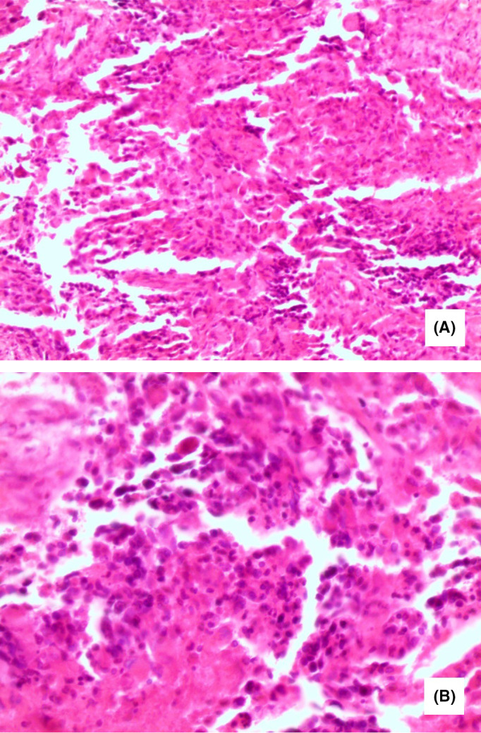
Pathology of the brain tumor showing: (A) Diffuse proliferation of variable sized anaplastic cells occurring in small groups and isolated forms with large round or elongated hyperchromatic nuclei, and occasional bizarre mitotic figures (H&E ×200). B, Higher magnification with scattered foci of necrosis (H&E ×400)
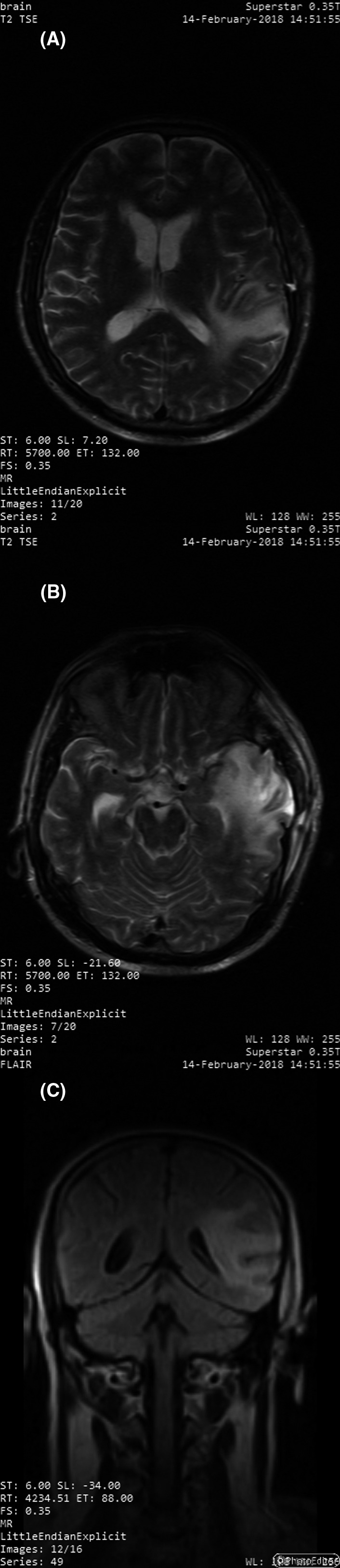
MRI 4 mo after the surgery showing the area of the lesion with residual edema with no midline deviation nor ventricular compression on transverse T2 TSE images (A and B) and coronal FLAIR image (C)
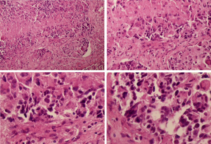
Pathological microscopic images of the lymph node biopsy showing: invasive proliferating anaplastic cells, large hyperchromatic nuclei and occasional bizarre mitotic figures, with no identified residual lymphoid tissue
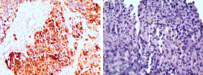
Immune staining of the lymph node biopsy confirming the GBM origin with positive GFAP staining (on the left) and negative CK staining (on the right)
References
-
- Louis DN, Ohgaki H, Wiestler OD, Cavenee WK. WHO Classification of Tumours of the Central Nervous System, Revised (4th edn). WHO, WHO Press, Geneva, Switzerland: WHO ‐ OMS. Fourth. IARC; 2016. http://apps.who.int/bookorders/anglais/detart1.jsp?codlan=1&codcol=70&co...
Publication types
LinkOut - more resources
Full Text Sources
