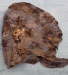
Left hypoplastic lung and hemoptysis-rare familial unilateral pulmonary vein atresia
- PMID: 32983480
- PMCID: PMC7495865
- DOI: 10.1002/ccr3.2982
Left hypoplastic lung and hemoptysis-rare familial unilateral pulmonary vein atresia
Abstract
Unilateral pulmonary vein atresia (UPVA) is a rare congenital vascular malformation with obliteration of the pulmonary vein. We present a case series of three siblings with variable presentation of UPVA. We suggest a dominant genetic cause based on different paternity. Identifying genetic etiology would contribute to early diagnosis and screening.
Keywords: pulmonary hypertension (PH); pulmonary vein stenosis (PVS); unilateral pulmonary vein atresia (UPVA).
© 2020 The Authors. Clinical Case Reports published by John Wiley & Sons Ltd.
Conflict of interest statement
Dr Cohn, Dr A Hicks, Dr Lacson, and Dr M Hicks declare that they have no competing interests and did not receive any personal financial support.
Figures


Similar articles
-
Unilateral pulmonary vein atresia.IJTLD Open. 2025 Apr 9;2(4):224-229. doi: 10.5588/ijtldopen.24.0631. eCollection 2025 Apr. IJTLD Open. 2025. PMID: 40226141 Free PMC article.
-
Unilateral pulmonary vein atresia presenting with recurrent hydrothorax in an adult: A case report.Respir Med Case Rep. 2022 Aug 13;39:101711. doi: 10.1016/j.rmcr.2022.101711. eCollection 2022. Respir Med Case Rep. 2022. PMID: 36060639 Free PMC article.
-
Unilateral pulmonary vein atresia without anomalous connection in adult patient with recurrent severe hemoptysis.J Vis Surg. 2018 May 22;4:111. doi: 10.21037/jovs.2018.05.03. eCollection 2018. J Vis Surg. 2018. PMID: 29963400 Free PMC article.
-
Isolated unilateral pulmonary vein atresia with hemoptysis in a child: A case report and literature review.Medicine (Baltimore). 2018 Aug;97(34):e11882. doi: 10.1097/MD.0000000000011882. Medicine (Baltimore). 2018. PMID: 30142786 Free PMC article. Review.
-
Isolated unilateral pulmonary vein atresia in adult patients: a case report and literature review.Heart Surg Forum. 2010 Dec;13(6):E370-2. doi: 10.1532/HSF98.20101078. Heart Surg Forum. 2010. PMID: 21169144 Review.
Cited by
-
Unilateral pulmonary vein atresia.IJTLD Open. 2025 Apr 9;2(4):224-229. doi: 10.5588/ijtldopen.24.0631. eCollection 2025 Apr. IJTLD Open. 2025. PMID: 40226141 Free PMC article.
-
Unilateral pulmonary vein atresia presenting with recurrent hemoptysis and bronchial varices in an Ethiopian adolescent: a case report.J Med Case Rep. 2023 Jun 3;17(1):246. doi: 10.1186/s13256-023-03956-4. J Med Case Rep. 2023. PMID: 37269023 Free PMC article.
-
Prematurity and Pulmonary Vein Stenosis: The Role of Parenchymal Lung Disease and Pulmonary Vascular Disease.Children (Basel). 2022 May 12;9(5):713. doi: 10.3390/children9050713. Children (Basel). 2022. PMID: 35626890 Free PMC article. Review.
References
-
- Kendig's Disorders of the Respiratory Tract in Children (9th edn Philadelphia: Elsevier; ) 2019;(384):326‐327.
-
- Stillwell PC, Kupfer O. Hemoptysis in children. UpToDate. 2017;1‐8.
-
- Hull J, Julian F, Anne T. Hemoptysis Pediatric respiratory Medicine, Oxford Specialist Handbooks in Pediatrics. , (2nd edn). Oxford, UK: Oxford University Press; 2015:61‐65.
-
- Lara AR, Schwarz MI. Diffuse alveolar hemorrhage. Chest. 2010;137(5):1164‐1171. - PubMed
-
- Brasher P, Klein RJ, Fantauzzi J, Judson MA, Chopra A. A 23y old man with recurrent hemoptysis. Pulmonary Critical care and sleep pearls. Chest. 2015;148(5):152‐155. - PubMed
Publication types
LinkOut - more resources
Full Text Sources
Research Materials

