

Case Reports
doi: 10.1002/ccr3.2953.
eCollection 2020 Sep.
A Japanese case of amoebic meningoencephalitis initially diagnosed by cerebrospinal fluid cytology
Affiliations
- PMID: 32983486
- PMCID: PMC7495867
- DOI: 10.1002/ccr3.2953
Item in Clipboard
Case Reports
A Japanese case of amoebic meningoencephalitis initially diagnosed by cerebrospinal fluid cytology
Clin Case Rep.
.
Abstract
Microscopy can detect the presence of amoebic trophozoites in cerebrospinal fluid and tissue. The infection was confirmed in the present case by polymerase chain reaction and immunohistochemistry, but we were unable to achieve a cure. Our case rapidly progressed without any skin lesions.
Keywords: Balamuthia mandrillaris amoebic meningoencephalitis; Free‐living amoebae; PCR; cerebrospinal fluid; cytology; immunohistochemistry.
© 2020 The Authors. Clinical Case Reports published by John Wiley & Sons Ltd.
Conflict of interest statement
None declared.
Figures

Follow‐up MRI after lung cancer surgery. A, Axial T1‐weighted MRI shows a ring‐enhancing lesion in the right occipital lobe, suggesting brain metastasis of lung cancer. B, Six months later, MRI shows that the lesion has grown despite stereotactic radiosurgery
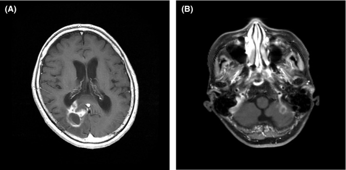
Three months after the removal of the brain lesion, axial T1‐weighted MRI shows (A) epidural and endocranial invasion of the lesion and (B) a new lesion that has developed in the left cerebellar hemisphere

Amoebic trophozoites present in the cerebrospinal fluid. Note the oval or round shape of the trophozoites with mono‐ (A) and multiple (B, C) nuclei (Sternheimer's stain, original magnification ×100)

(A‐C) Many macrophages and protozoa‐like structures with mono‐ or multiple nuclei can be seen in the cerebrospinal fluid, findings that are suggestive of amoebic meningoencephalitis (Papanicolaou's stain, bars = 10 µm)

A few atypical glandular cells suggestive of adenocarcinoma are present in the cerebrospinal fluid cytology (Papanicolaou's stain, bar = 10 µm)

The nasal discharge contains many amoebic trophozoites that are actively moving. Sternheimer's stain (original magnification, ×100)

Histopathology of the brain lesion. A, Amoebic trophozoites are present in the degenerated tissue. B, They are positive for PAS reaction. A, Hematoxylin and eosin (H&E) stain and B, Periodic acid‐Schiff (PAS) reaction, bars = 10 µm

Immunohistochemical stains using rabbit antisera against A, Acanthamoeba spp. and B, B mandrillaris for the brain lesion. The reaction is negative for Acanthamoeba spp. and positive for B mandrillaris. bars = 20 µm

An 18S rRNA gene polymerase chain reaction assay of formalin‐fixed, paraffin‐embedded brain‐tissue specimens showing the presence of Balamuthia DNA (150 bp). Line 1, 100‐bp molecular ladder (size marker); Lanes 2‐5, DNA amplification products from four different brain samples; Lane 6, Balamuthia‐positive control; and Lane 7, Balamuthia‐negative control (Milli Q water)
References
-
- Hara T, Yagita K, Sugita Y. Pathogenic free‐living amoebic encephalitis in Japan. Neuropathology. 2019;39(4):251‐258. - PubMed
-
- Visvesvara GS, Moura H, Schuster FL. Pathogenic and opportunistic free‐living amoebae: Acanthamoeba spp., Balamuthia mandrillaris, Naegleria fowleri, and Sappinia diploidea . FEMS Immunol Med Microbiol. 2007;50(1):1‐26. - PubMed
-
- Bando Y, Takahashi T, Uehara H, Kagegi T, Nagahiro S, Izumi K. Autopsy case of amebic granulomatous meningoencephalitis caused by Balamuthia mandrillaris in Japan. Pathol Int. 2012;62(6):418‐423. - PubMed
Publication types
LinkOut - more resources
Full Text Sources
