Diagnosis and Surgical Management of Exophytic Suprasellar Pituitary Adenoma Extending Over the Diaphragma Sellae and Mimicking Craniopharyngioma: A Case Report
- PMID: 32983721
- PMCID: PMC7515809
- DOI: 10.7759/cureus.10028
Diagnosis and Surgical Management of Exophytic Suprasellar Pituitary Adenoma Extending Over the Diaphragma Sellae and Mimicking Craniopharyngioma: A Case Report
Abstract
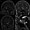
Pituitary adenomas developing from the lateral surface of the pituitary gland are referred to as exophytic pituitary adenomas. When an exophytic pituitary adenoma extends into the suprasellar region, the tumor exhibits an atypical growth pattern that makes it difficult to distinguish it from craniopharyngiomas or other parasellar lesions on MRI. A 53-year-old woman who presented with general malaise and visual disturbances was diagnosed with a brain tumor. MRI showed a suprasellar tumor presenting as superior lobulation with reticular enhancement and partial calcification. Subsequently, endoscopic transsphenoidal surgery was performed on the patient. The suprasellar tumor was found to originate from the superior surface of the normal pituitary gland and it extended into the supra-diaphragm region. Subtotal tumor resection was achieved, and her clinical symptoms subsequently improved. Exophytic suprasellar pituitary adenomas (SPAs) are extremely rare and may be mistaken for ectopic SPAs in some cases. Contrast-enhanced fast imaging employing steady-state acquisition (CE-FIESTA) can clearly depict the connection between an exophytic SPA and the normal pituitary gland via a diaphragma sellae defect. During surgery, it was seen that the exophytic SPA and anterior lobe of the pituitary gland connected with each other directly. The tumor originated from the superior surface of the pituitary gland and extended into the supra-diaphragm region. These findings clearly confirmed the difference between exophytic SPAs and ectopic SPAs. In surgical management, an exophytic SPA needs careful consideration for resecting the tumor from encased surrounding structures without vascular and nerve injury. Ultrasonic aspiration devices may be useful for safely resecting the tumor from important structures due to tissue selection. Exophytic SPAs are distinguished from ectopic SPAs with respect to the direct connection between the tumor and the normal pituitary gland. These findings can be clearly depicted using CE-FIESTA and should be confirmed during surgery. Clinicians should be aware of the risk that exophytic SPA may extend into the supra-diaphragm region and of the difficulties of resecting the tumor encasing surrounding structures in the suprasellar region.
Keywords: craniopharyngioma; ectopic pituitary adenoma; endoscopic transsphenidal surgery; exophytic pituitary adenoma; fiesta; silent corticotroph adenoma; suprasellar pituitary adenoma; ultrasonic aspiration.
Copyright © 2020, Noguchi et al.
Conflict of interest statement
The authors have declared that no competing interests exist.
Figures


Similar articles
-
Suprasellar pituitary adenomas: a 10-year experience in a single tertiary medical center and a literature review.Pituitary. 2020 Aug;23(4):367-380. doi: 10.1007/s11102-020-01043-1. Pituitary. 2020. PMID: 32378170 Review.
-
'Ectopic' suprasellar type IIa PRL-secreting pituitary adenoma.Pituitary. 2017 Aug;20(4):477-484. doi: 10.1007/s11102-017-0807-9. Pituitary. 2017. PMID: 28526958 Review.
-
Extended endoscopic endonasal surgery using three-dimensional endoscopy in the intra-operative MRI suite for supra-diaphragmatic ectopic pituitary adenoma.Turk Neurosurg. 2015;25(3):503-7. doi: 10.5137/1019-5149.JTN.11212-14.1. Turk Neurosurg. 2015. PMID: 26037197
-
[Suprasellar ectopic pituitary adenoma: report of a case].No Shinkei Geka. 1994 Dec;22(12):1141-5. No Shinkei Geka. 1994. PMID: 7845510 Review. Japanese.
-
[Cystic ectopic pituitary adenoma: report of a case].No To Shinkei. 1995 Nov;47(11):1092-7. No To Shinkei. 1995. PMID: 7495616 Review. Japanese.
Cited by
-
Clinical and radiological presentation of parasellar ectopic pituitary adenomas: case series and systematic review of the literature.J Endocrinol Invest. 2022 Aug;45(8):1465-1481. doi: 10.1007/s40618-022-01758-x. Epub 2022 Feb 11. J Endocrinol Invest. 2022. PMID: 35147925
-
Calcified Pituitary Adenoma Mimicking Craniopharyngioma: A Case Report.Cureus. 2024 Feb 17;16(2):e54352. doi: 10.7759/cureus.54352. eCollection 2024 Feb. Cureus. 2024. PMID: 38500912 Free PMC article.
References
-
- Pituitary adenoma, craniopharyngioma, and Rathke cleft cyst involving both intrasellar and suprasellar regions: differentiation using MRI. Choi SH, Kwon BJ, Na DG, Kim JH, Han MH, Chang KH. Clin Radiol. 2007;62:453–462. - PubMed
-
- Directional regulation of extrasellar extension by sellar dura integrity and intrasphenoidal septation in pituitary adenomas. Hayashi Y, Sasagawa Y, Oishi M, et al. World Neurosurg. 2019;122:0. - PubMed
-
- Outcomes of three-Tesla magnetic resonance imaging for the identification of pituitary adenoma in patients with Cushing's disease. Fukuhara N, Inoshita N, Yamaguchi-Okada M, et al. Endocr J. 2019;66:259–264. - PubMed
-
- Surgical management of pituitary tumors. Wilson CB. J Clin Endocrinol Metab. 1997;82:2381–2385. - PubMed
-
- Microsurgical anatomy of the diaphragma sellae and its role in directing the pattern of growth of pituitary adenomas. Campero A, Martins C, Yasuda A, Rhoton AL Jr. Neurosurgery. 2008;62:717–723. - PubMed
Publication types
LinkOut - more resources
Full Text Sources
